Calcium »
PDB 5npf-5odc »
5nrk »
Calcium in PDB 5nrk: Crystal Structure of the Sixth Cohesin From Acetivibrio Cellulolyticus' Scaffoldin B in Complex with CEL5 Dockerin S15I, I16N Mutant
Enzymatic activity of Crystal Structure of the Sixth Cohesin From Acetivibrio Cellulolyticus' Scaffoldin B in Complex with CEL5 Dockerin S15I, I16N Mutant
All present enzymatic activity of Crystal Structure of the Sixth Cohesin From Acetivibrio Cellulolyticus' Scaffoldin B in Complex with CEL5 Dockerin S15I, I16N Mutant:
3.2.1.4;
3.2.1.4;
Protein crystallography data
The structure of Crystal Structure of the Sixth Cohesin From Acetivibrio Cellulolyticus' Scaffoldin B in Complex with CEL5 Dockerin S15I, I16N Mutant, PDB code: 5nrk
was solved by
P.Bule,
S.Najmudin,
C.M.G.A.Fontes,
V.D.Alves,
with X-Ray Crystallography technique. A brief refinement statistics is given in the table below:
| Resolution Low / High (Å) | 65.03 / 1.45 |
| Space group | P 21 21 21 |
| Cell size a, b, c (Å), α, β, γ (°) | 46.539, 79.809, 112.159, 90.00, 90.00, 90.00 |
| R / Rfree (%) | 14.4 / 17 |
Calcium Binding Sites:
The binding sites of Calcium atom in the Crystal Structure of the Sixth Cohesin From Acetivibrio Cellulolyticus' Scaffoldin B in Complex with CEL5 Dockerin S15I, I16N Mutant
(pdb code 5nrk). This binding sites where shown within
5.0 Angstroms radius around Calcium atom.
In total 6 binding sites of Calcium where determined in the Crystal Structure of the Sixth Cohesin From Acetivibrio Cellulolyticus' Scaffoldin B in Complex with CEL5 Dockerin S15I, I16N Mutant, PDB code: 5nrk:
Jump to Calcium binding site number: 1; 2; 3; 4; 5; 6;
In total 6 binding sites of Calcium where determined in the Crystal Structure of the Sixth Cohesin From Acetivibrio Cellulolyticus' Scaffoldin B in Complex with CEL5 Dockerin S15I, I16N Mutant, PDB code: 5nrk:
Jump to Calcium binding site number: 1; 2; 3; 4; 5; 6;
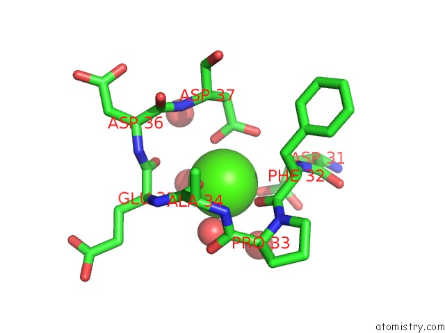
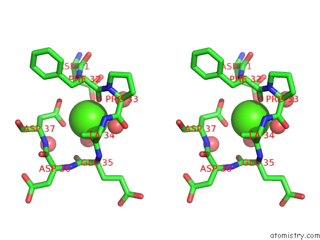
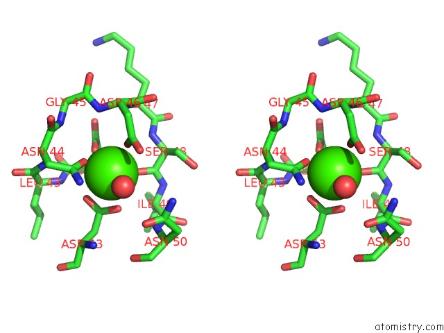
Calcium binding site 1 out of 6 in 5nrk
Go back to
Calcium binding site 1 out
of 6 in the Crystal Structure of the Sixth Cohesin From Acetivibrio Cellulolyticus' Scaffoldin B in Complex with CEL5 Dockerin S15I, I16N Mutant
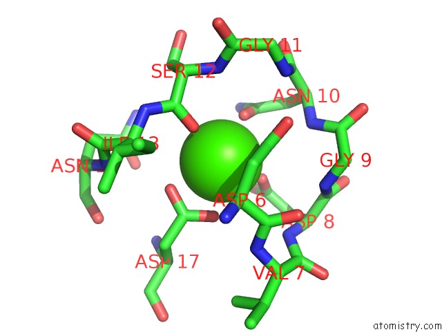
Mono view
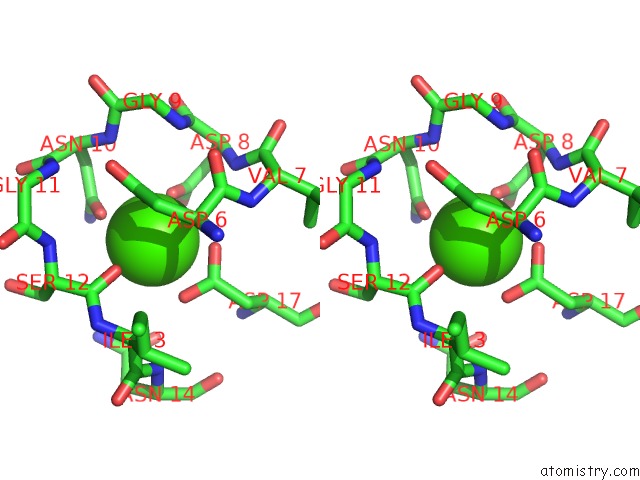
Stereo pair view
Mono view
Stereo pair view
A full contact list of Calcium with other atoms in the Ca binding
site number 1 of Crystal Structure of the Sixth Cohesin From Acetivibrio Cellulolyticus' Scaffoldin B in Complex with CEL5 Dockerin S15I, I16N Mutant within 5.0Å range:
|
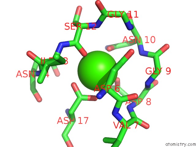
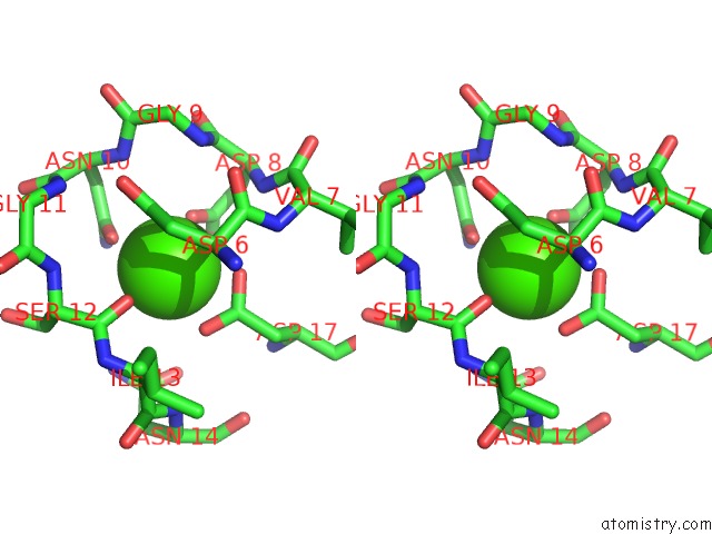
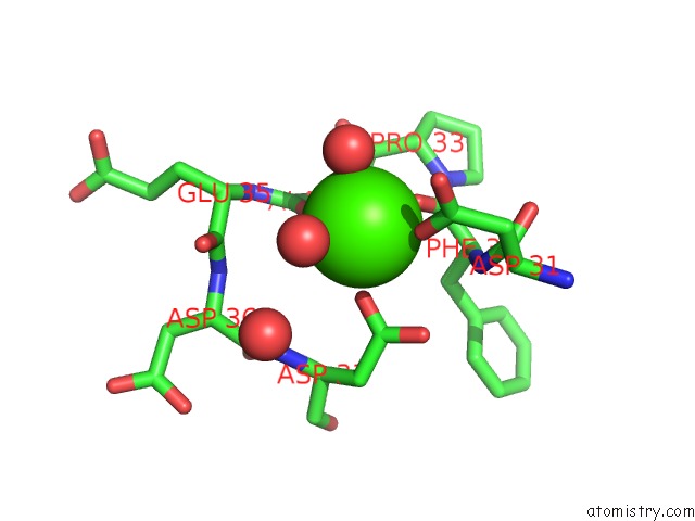
Calcium binding site 2 out of 6 in 5nrk
Go back to
Calcium binding site 2 out
of 6 in the Crystal Structure of the Sixth Cohesin From Acetivibrio Cellulolyticus' Scaffoldin B in Complex with CEL5 Dockerin S15I, I16N Mutant
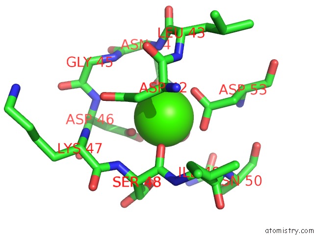
Mono view
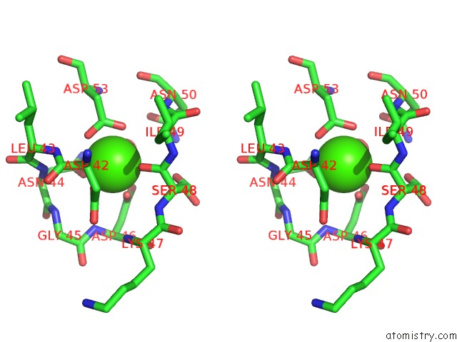
Stereo pair view
Mono view
Stereo pair view
A full contact list of Calcium with other atoms in the Ca binding
site number 2 of Crystal Structure of the Sixth Cohesin From Acetivibrio Cellulolyticus' Scaffoldin B in Complex with CEL5 Dockerin S15I, I16N Mutant within 5.0Å range:
|
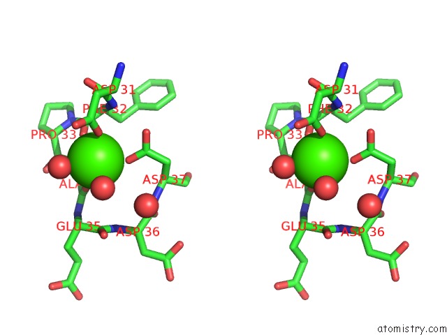
Calcium binding site 3 out of 6 in 5nrk
Go back to
Calcium binding site 3 out
of 6 in the Crystal Structure of the Sixth Cohesin From Acetivibrio Cellulolyticus' Scaffoldin B in Complex with CEL5 Dockerin S15I, I16N Mutant
Mono view
Stereo pair view
Mono view
Stereo pair view
A full contact list of Calcium with other atoms in the Ca binding
site number 3 of Crystal Structure of the Sixth Cohesin From Acetivibrio Cellulolyticus' Scaffoldin B in Complex with CEL5 Dockerin S15I, I16N Mutant within 5.0Å range:
|
Calcium binding site 4 out of 6 in 5nrk
Go back to
Calcium binding site 4 out
of 6 in the Crystal Structure of the Sixth Cohesin From Acetivibrio Cellulolyticus' Scaffoldin B in Complex with CEL5 Dockerin S15I, I16N Mutant
Mono view
Stereo pair view
Mono view
Stereo pair view
A full contact list of Calcium with other atoms in the Ca binding
site number 4 of Crystal Structure of the Sixth Cohesin From Acetivibrio Cellulolyticus' Scaffoldin B in Complex with CEL5 Dockerin S15I, I16N Mutant within 5.0Å range:
|
Calcium binding site 5 out of 6 in 5nrk
Go back to
Calcium binding site 5 out
of 6 in the Crystal Structure of the Sixth Cohesin From Acetivibrio Cellulolyticus' Scaffoldin B in Complex with CEL5 Dockerin S15I, I16N Mutant

Mono view
Stereo pair view
Mono view
Stereo pair view
A full contact list of Calcium with other atoms in the Ca binding
site number 5 of Crystal Structure of the Sixth Cohesin From Acetivibrio Cellulolyticus' Scaffoldin B in Complex with CEL5 Dockerin S15I, I16N Mutant within 5.0Å range:
|
Calcium binding site 6 out of 6 in 5nrk
Go back to
Calcium binding site 6 out
of 6 in the Crystal Structure of the Sixth Cohesin From Acetivibrio Cellulolyticus' Scaffoldin B in Complex with CEL5 Dockerin S15I, I16N Mutant
Mono view
Stereo pair view
Mono view
Stereo pair view
A full contact list of Calcium with other atoms in the Ca binding
site number 6 of Crystal Structure of the Sixth Cohesin From Acetivibrio Cellulolyticus' Scaffoldin B in Complex with CEL5 Dockerin S15I, I16N Mutant within 5.0Å range:
|
Reference:
P.Bule,
K.Cameron,
J.A.M.Prates,
L.M.A.Ferreira,
S.P.Smith,
H.J.Gilbert,
E.A.Bayer,
S.Najmudin,
C.M.G.A.Fontes,
V.D.Alves.
Structure-Function Analyses Generate Novel Specificities to Assemble the Components of Multienzyme Bacterial Cellulosome Complexes. J. Biol. Chem. V. 293 4201 2018.
ISSN: ESSN 1083-351X
PubMed: 29367338
DOI: 10.1074/JBC.RA117.001241
Page generated: Wed Jul 9 09:08:33 2025
ISSN: ESSN 1083-351X
PubMed: 29367338
DOI: 10.1074/JBC.RA117.001241
Last articles
Mg in 1N5KMg in 1N5J
Mg in 1N56
Mg in 1N57
Mg in 1N52
Mg in 1N2C
Mg in 1N24
Mg in 1N2G
Mg in 1N2S
Mg in 1N23