Calcium »
PDB 5npf-5odc »
5o8l »
Calcium in PDB 5o8l: Crystal Structure of Leuconostoc Citreum Nrrl B-1299 N-Terminally Truncated Dextransucrase Dsr-M in Complex with Sucrose
Enzymatic activity of Crystal Structure of Leuconostoc Citreum Nrrl B-1299 N-Terminally Truncated Dextransucrase Dsr-M in Complex with Sucrose
All present enzymatic activity of Crystal Structure of Leuconostoc Citreum Nrrl B-1299 N-Terminally Truncated Dextransucrase Dsr-M in Complex with Sucrose:
2.4.1.140;
2.4.1.140;
Protein crystallography data
The structure of Crystal Structure of Leuconostoc Citreum Nrrl B-1299 N-Terminally Truncated Dextransucrase Dsr-M in Complex with Sucrose, PDB code: 5o8l
was solved by
M.Claverie,
G.Cioci,
M.Remaud-Simeon,
C.Moulis,
S.Tranier,
with X-Ray Crystallography technique. A brief refinement statistics is given in the table below:
| Resolution Low / High (Å) | 50.00 / 3.60 |
| Space group | P 31 2 1 |
| Cell size a, b, c (Å), α, β, γ (°) | 183.625, 183.625, 146.804, 90.00, 90.00, 120.00 |
| R / Rfree (%) | 19.8 / 22.8 |
Calcium Binding Sites:
The binding sites of Calcium atom in the Crystal Structure of Leuconostoc Citreum Nrrl B-1299 N-Terminally Truncated Dextransucrase Dsr-M in Complex with Sucrose
(pdb code 5o8l). This binding sites where shown within
5.0 Angstroms radius around Calcium atom.
In total only one binding site of Calcium was determined in the Crystal Structure of Leuconostoc Citreum Nrrl B-1299 N-Terminally Truncated Dextransucrase Dsr-M in Complex with Sucrose, PDB code: 5o8l:
In total only one binding site of Calcium was determined in the Crystal Structure of Leuconostoc Citreum Nrrl B-1299 N-Terminally Truncated Dextransucrase Dsr-M in Complex with Sucrose, PDB code: 5o8l:
Calcium binding site 1 out of 1 in 5o8l
Go back to
Calcium binding site 1 out
of 1 in the Crystal Structure of Leuconostoc Citreum Nrrl B-1299 N-Terminally Truncated Dextransucrase Dsr-M in Complex with Sucrose
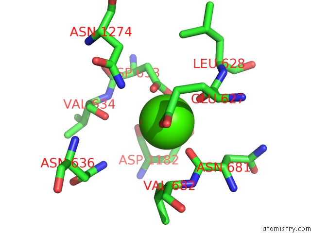
Mono view
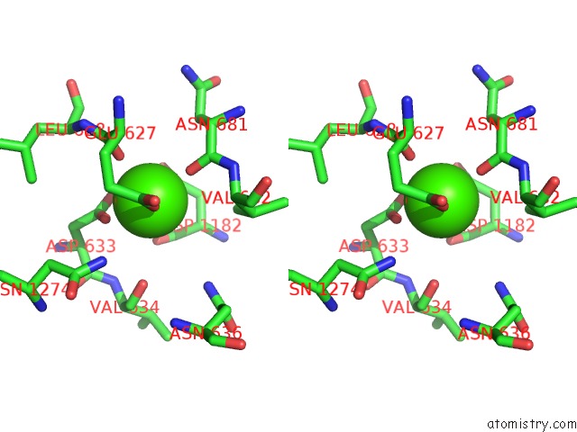
Stereo pair view
Mono view
Stereo pair view
A full contact list of Calcium with other atoms in the Ca binding
site number 1 of Crystal Structure of Leuconostoc Citreum Nrrl B-1299 N-Terminally Truncated Dextransucrase Dsr-M in Complex with Sucrose within 5.0Å range:
|
Reference:
M.Claverie,
G.Cioci,
M.Vuillemin,
N.Monties,
P.Roblin,
G.Lippens,
M.Remaud-Simeon,
C.Moulis.
Investigations on the Determinants Responsible For Low Molar Mass Dextran Formation By Dsr-M Dextransucrase Acs Catalysis V. 7 7106 2017.
ISSN: ESSN 2155-5435
DOI: 10.1021/ACSCATAL.7B02182
Page generated: Wed Jul 9 09:15:03 2025
ISSN: ESSN 2155-5435
DOI: 10.1021/ACSCATAL.7B02182
Last articles
Mg in 1X9WMg in 1X9S
Mg in 1X8B
Mg in 1X9M
Mg in 1X84
Mg in 1X83
Mg in 1X88
Mg in 1X55
Mg in 1X54
Mg in 1X3S