Calcium »
PDB 5npf-5odc »
5o8x »
Calcium in PDB 5o8x: The X-Ray Structure of Catenated Lytic Transglycosylase SLTB1 From Pseudomonas Aeruginosa
Protein crystallography data
The structure of The X-Ray Structure of Catenated Lytic Transglycosylase SLTB1 From Pseudomonas Aeruginosa, PDB code: 5o8x
was solved by
T.Dominguez-Gil,
R.Molina,
with X-Ray Crystallography technique. A brief refinement statistics is given in the table below:
| Resolution Low / High (Å) | 48.26 / 2.50 |
| Space group | C 1 2 1 |
| Cell size a, b, c (Å), α, β, γ (°) | 115.064, 116.358, 54.865, 90.00, 118.41, 90.00 |
| R / Rfree (%) | 17.7 / 22.9 |
Other elements in 5o8x:
The structure of The X-Ray Structure of Catenated Lytic Transglycosylase SLTB1 From Pseudomonas Aeruginosa also contains other interesting chemical elements:
| Arsenic | (As) | 2 atoms |
Calcium Binding Sites:
The binding sites of Calcium atom in the The X-Ray Structure of Catenated Lytic Transglycosylase SLTB1 From Pseudomonas Aeruginosa
(pdb code 5o8x). This binding sites where shown within
5.0 Angstroms radius around Calcium atom.
In total 2 binding sites of Calcium where determined in the The X-Ray Structure of Catenated Lytic Transglycosylase SLTB1 From Pseudomonas Aeruginosa, PDB code: 5o8x:
Jump to Calcium binding site number: 1; 2;
In total 2 binding sites of Calcium where determined in the The X-Ray Structure of Catenated Lytic Transglycosylase SLTB1 From Pseudomonas Aeruginosa, PDB code: 5o8x:
Jump to Calcium binding site number: 1; 2;
Calcium binding site 1 out of 2 in 5o8x
Go back to
Calcium binding site 1 out
of 2 in the The X-Ray Structure of Catenated Lytic Transglycosylase SLTB1 From Pseudomonas Aeruginosa
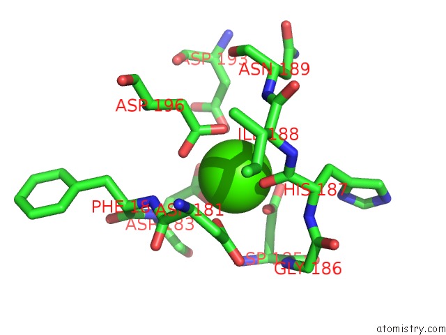
Mono view
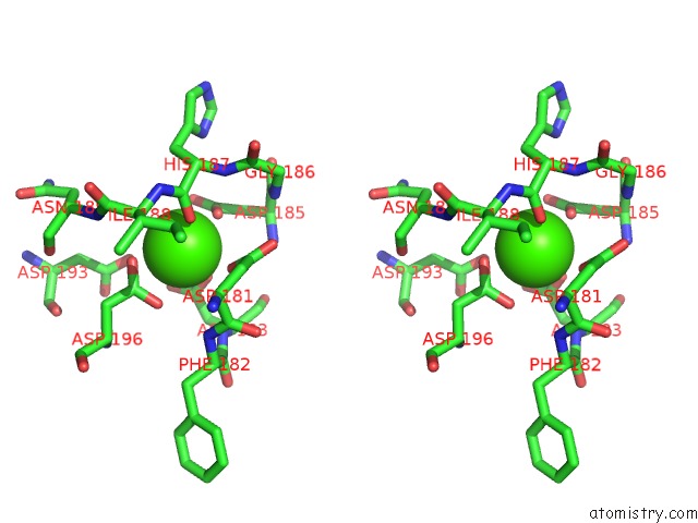
Stereo pair view
Mono view
Stereo pair view
A full contact list of Calcium with other atoms in the Ca binding
site number 1 of The X-Ray Structure of Catenated Lytic Transglycosylase SLTB1 From Pseudomonas Aeruginosa within 5.0Å range:
|
Calcium binding site 2 out of 2 in 5o8x
Go back to
Calcium binding site 2 out
of 2 in the The X-Ray Structure of Catenated Lytic Transglycosylase SLTB1 From Pseudomonas Aeruginosa
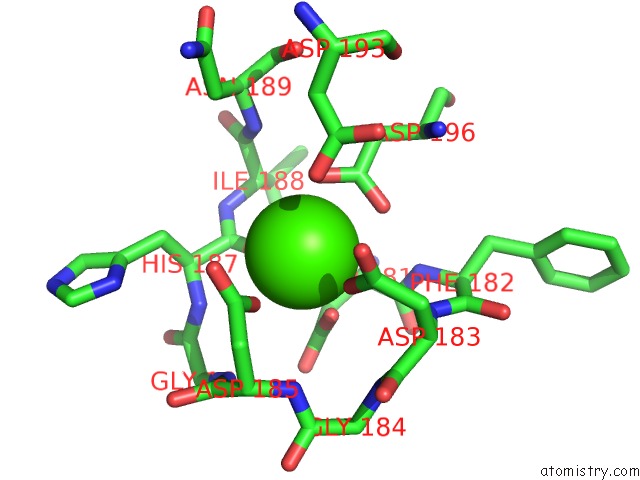
Mono view
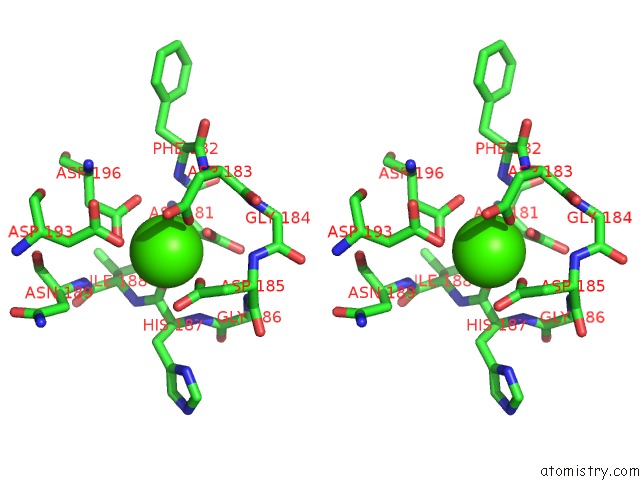
Stereo pair view
Mono view
Stereo pair view
A full contact list of Calcium with other atoms in the Ca binding
site number 2 of The X-Ray Structure of Catenated Lytic Transglycosylase SLTB1 From Pseudomonas Aeruginosa within 5.0Å range:
|
Reference:
T.Dominguez-Gil,
R.Molina,
D.A.Dik,
E.Spink,
S.Mobashery,
J.A.Hermoso.
X-Ray Structure of Catenated Lytic Transglycosylase SLTB1. Biochemistry V. 56 6317 2017.
ISSN: ISSN 1520-4995
PubMed: 29131935
DOI: 10.1021/ACS.BIOCHEM.7B00932
Page generated: Wed Jul 9 09:15:24 2025
ISSN: ISSN 1520-4995
PubMed: 29131935
DOI: 10.1021/ACS.BIOCHEM.7B00932
Last articles
K in 8I03K in 8IAT
K in 8I02
K in 8HZF
K in 8HZM
K in 8HZJ
K in 8HKQ
K in 8HPO
K in 8HZE
K in 8HK6