Calcium »
PDB 5odu-5oyl »
5oyl »
Calcium in PDB 5oyl: Vsv G CR2
Protein crystallography data
The structure of Vsv G CR2, PDB code: 5oyl
was solved by
A.A.Albertini,
L.Belot,
P.Legrand,
Y.Gaudin,
with X-Ray Crystallography technique. A brief refinement statistics is given in the table below:
| Resolution Low / High (Å) | 29.98 / 2.25 |
| Space group | H 3 2 |
| Cell size a, b, c (Å), α, β, γ (°) | 90.040, 90.040, 515.780, 90.00, 90.00, 120.00 |
| R / Rfree (%) | 18.8 / 22.4 |
Calcium Binding Sites:
The binding sites of Calcium atom in the Vsv G CR2
(pdb code 5oyl). This binding sites where shown within
5.0 Angstroms radius around Calcium atom.
In total 2 binding sites of Calcium where determined in the Vsv G CR2, PDB code: 5oyl:
Jump to Calcium binding site number: 1; 2;
In total 2 binding sites of Calcium where determined in the Vsv G CR2, PDB code: 5oyl:
Jump to Calcium binding site number: 1; 2;
Calcium binding site 1 out of 2 in 5oyl
Go back to
Calcium binding site 1 out
of 2 in the Vsv G CR2
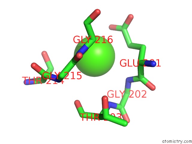
Mono view
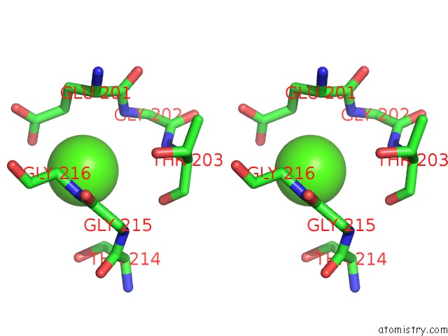
Stereo pair view
Mono view
Stereo pair view
A full contact list of Calcium with other atoms in the Ca binding
site number 1 of Vsv G CR2 within 5.0Å range:
|
Calcium binding site 2 out of 2 in 5oyl
Go back to
Calcium binding site 2 out
of 2 in the Vsv G CR2
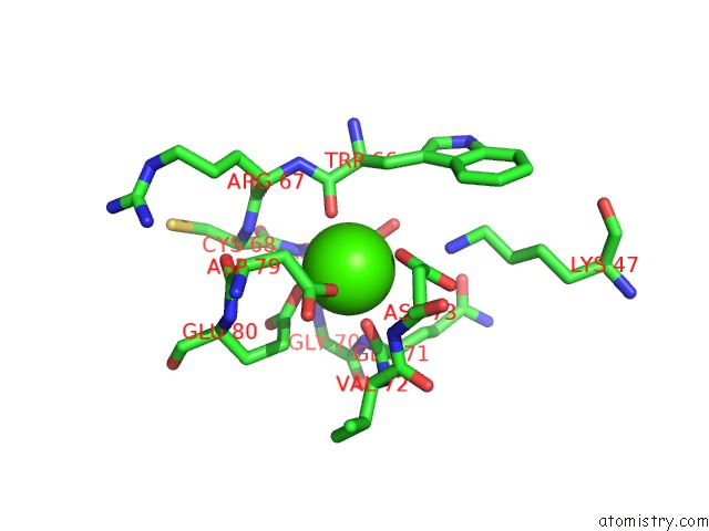
Mono view
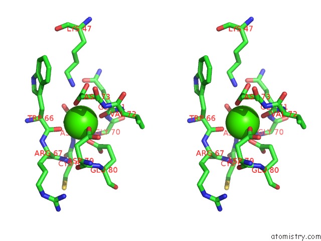
Stereo pair view
Mono view
Stereo pair view
A full contact list of Calcium with other atoms in the Ca binding
site number 2 of Vsv G CR2 within 5.0Å range:
|
Reference:
J.Nikolic,
L.Belot,
H.Raux,
P.Legrand,
Y.Gaudin,
A.A Albertini.
Structural Basis For the Recognition of Ldl-Receptor Family Members By Vsv Glycoprotein. Nat Commun V. 9 1029 2018.
ISSN: ESSN 2041-1723
PubMed: 29531262
DOI: 10.1038/S41467-018-03432-4
Page generated: Wed Jul 9 09:25:15 2025
ISSN: ESSN 2041-1723
PubMed: 29531262
DOI: 10.1038/S41467-018-03432-4
Last articles
Mg in 6CA4Mg in 6C90
Mg in 6CA0
Mg in 6C9Y
Mg in 6C8Z
Mg in 6C8P
Mg in 6C8N
Mg in 6C8O
Mg in 6C8D
Mg in 6C8L