Calcium »
PDB 5s5v-5szl »
5sv0 »
Calcium in PDB 5sv0: Structure of the Exbb/Exbd Complex From E. Coli at pH 7.0
Protein crystallography data
The structure of Structure of the Exbb/Exbd Complex From E. Coli at pH 7.0, PDB code: 5sv0
was solved by
H.Celia,
I.Botos,
R.Lloubes,
S.K.Buchanan,
N.Noinaj,
with X-Ray Crystallography technique. A brief refinement statistics is given in the table below:
| Resolution Low / High (Å) | 46.00 / 2.60 |
| Space group | P 1 21 1 |
| Cell size a, b, c (Å), α, β, γ (°) | 137.166, 104.779, 149.364, 90.00, 91.77, 90.00 |
| R / Rfree (%) | 21.1 / 25.8 |
Calcium Binding Sites:
The binding sites of Calcium atom in the Structure of the Exbb/Exbd Complex From E. Coli at pH 7.0
(pdb code 5sv0). This binding sites where shown within
5.0 Angstroms radius around Calcium atom.
In total 2 binding sites of Calcium where determined in the Structure of the Exbb/Exbd Complex From E. Coli at pH 7.0, PDB code: 5sv0:
Jump to Calcium binding site number: 1; 2;
In total 2 binding sites of Calcium where determined in the Structure of the Exbb/Exbd Complex From E. Coli at pH 7.0, PDB code: 5sv0:
Jump to Calcium binding site number: 1; 2;
Calcium binding site 1 out of 2 in 5sv0
Go back to
Calcium binding site 1 out
of 2 in the Structure of the Exbb/Exbd Complex From E. Coli at pH 7.0
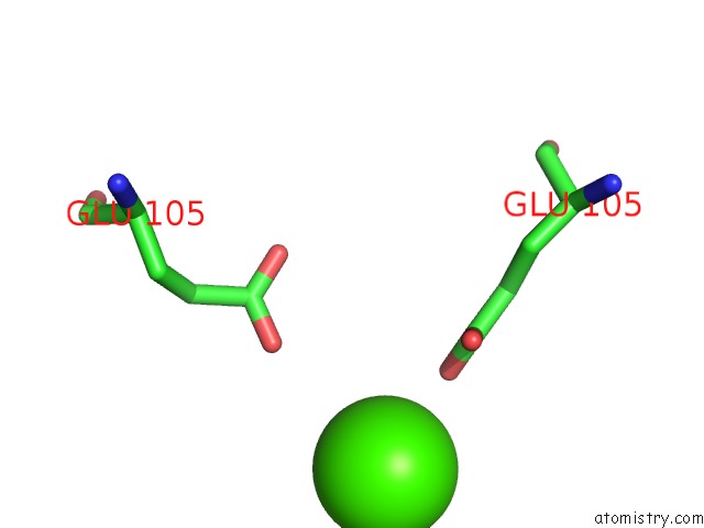
Mono view
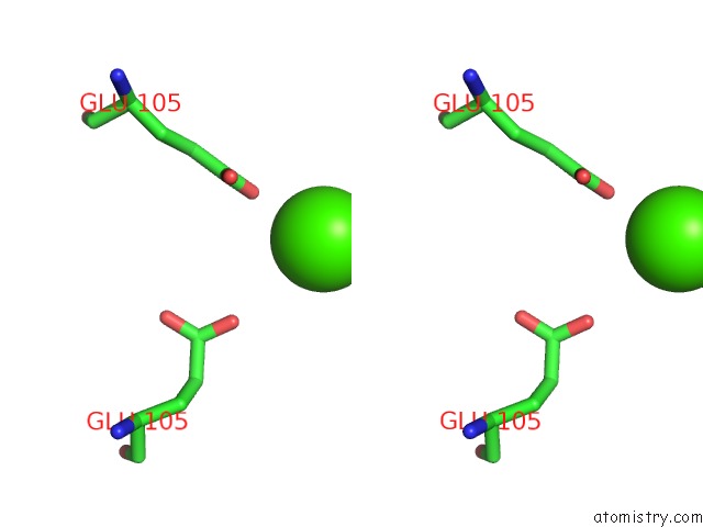
Stereo pair view
Mono view
Stereo pair view
A full contact list of Calcium with other atoms in the Ca binding
site number 1 of Structure of the Exbb/Exbd Complex From E. Coli at pH 7.0 within 5.0Å range:
|
Calcium binding site 2 out of 2 in 5sv0
Go back to
Calcium binding site 2 out
of 2 in the Structure of the Exbb/Exbd Complex From E. Coli at pH 7.0
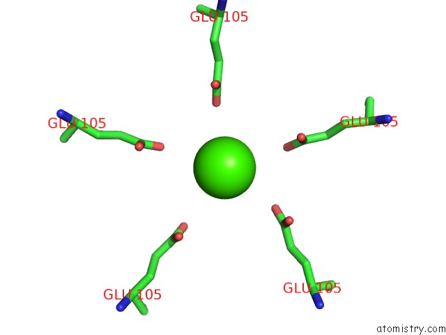
Mono view
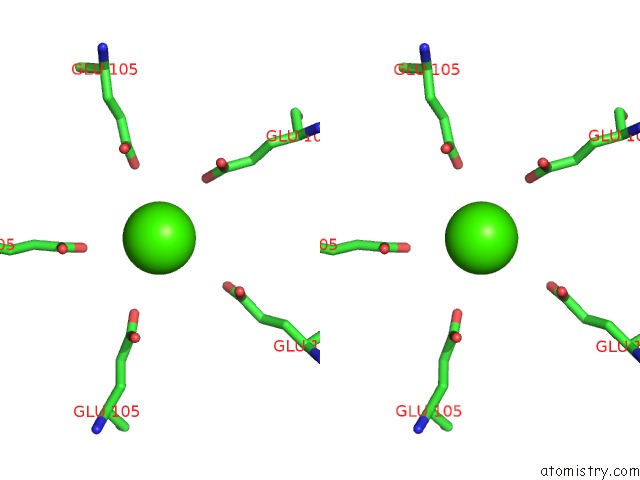
Stereo pair view
Mono view
Stereo pair view
A full contact list of Calcium with other atoms in the Ca binding
site number 2 of Structure of the Exbb/Exbd Complex From E. Coli at pH 7.0 within 5.0Å range:
|
Reference:
H.Celia,
N.Noinaj,
S.D.Zakharov,
E.Bordignon,
I.Botos,
M.Santamaria,
T.J.Barnard,
W.A.Cramer,
R.Lloubes,
S.K.Buchanan.
Structural Insight Into the Role of the Ton Complex in Energy Transduction. Nature V. 538 60 2016.
ISSN: ESSN 1476-4687
PubMed: 27654919
DOI: 10.1038/NATURE19757
Page generated: Wed Jul 9 09:50:04 2025
ISSN: ESSN 1476-4687
PubMed: 27654919
DOI: 10.1038/NATURE19757
Last articles
I in 7Z76I in 7YDX
I in 7XME
I in 7YUZ
I in 7XMK
I in 7XC1
I in 7WWN
I in 7X4U
I in 7W6B
I in 7VE3