Calcium »
PDB 5tae-5tua »
5tgq »
Calcium in PDB 5tgq: Restriction-Modification System Type II R.Swai, Dna Free
Protein crystallography data
The structure of Restriction-Modification System Type II R.Swai, Dna Free, PDB code: 5tgq
was solved by
B.W.Shen,
B.L.Stoddard,
with X-Ray Crystallography technique. A brief refinement statistics is given in the table below:
| Resolution Low / High (Å) | 50.01 / 1.88 |
| Space group | P 2 21 21 |
| Cell size a, b, c (Å), α, β, γ (°) | 48.386, 65.225, 67.567, 90.00, 90.00, 90.00 |
| R / Rfree (%) | 19.8 / 27.9 |
Other elements in 5tgq:
The structure of Restriction-Modification System Type II R.Swai, Dna Free also contains other interesting chemical elements:
| Chlorine | (Cl) | 3 atoms |
Calcium Binding Sites:
The binding sites of Calcium atom in the Restriction-Modification System Type II R.Swai, Dna Free
(pdb code 5tgq). This binding sites where shown within
5.0 Angstroms radius around Calcium atom.
In total only one binding site of Calcium was determined in the Restriction-Modification System Type II R.Swai, Dna Free, PDB code: 5tgq:
In total only one binding site of Calcium was determined in the Restriction-Modification System Type II R.Swai, Dna Free, PDB code: 5tgq:
Calcium binding site 1 out of 1 in 5tgq
Go back to
Calcium binding site 1 out
of 1 in the Restriction-Modification System Type II R.Swai, Dna Free
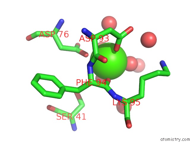
Mono view
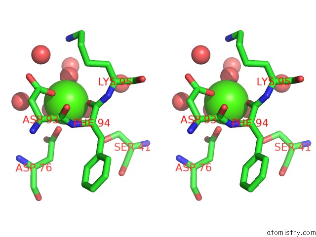
Stereo pair view
Mono view
Stereo pair view
A full contact list of Calcium with other atoms in the Ca binding
site number 1 of Restriction-Modification System Type II R.Swai, Dna Free within 5.0Å range:
|
Reference:
B.W.Shen,
D.F.Heiter,
K.D.Lunnen,
G.G.Wilson,
B.L.Stoddard.
Dna Recognition By the Swai Restriction Endonuclease Involves Unusual Distortion of An 8 Base Pair A:T-Rich Target. Nucleic Acids Res. V. 45 1516 2017.
ISSN: ESSN 1362-4962
PubMed: 28180307
DOI: 10.1093/NAR/GKW1200
Page generated: Wed Jul 9 10:17:57 2025
ISSN: ESSN 1362-4962
PubMed: 28180307
DOI: 10.1093/NAR/GKW1200
Last articles
Mn in 9LJUMn in 9LJW
Mn in 9LJS
Mn in 9LJR
Mn in 9LJT
Mn in 9LJV
Mg in 9UA2
Mg in 9R96
Mg in 9VM1
Mg in 9P01