Calcium »
PDB 5uwk-5vlk »
5veq »
Calcium in PDB 5veq: Mouse Kynurenine Aminotransferase III, Re-Refinement of the Pdb Structure 3E2Y
Enzymatic activity of Mouse Kynurenine Aminotransferase III, Re-Refinement of the Pdb Structure 3E2Y
All present enzymatic activity of Mouse Kynurenine Aminotransferase III, Re-Refinement of the Pdb Structure 3E2Y:
2.6.1.63; 2.6.1.7; 4.4.1.13;
2.6.1.63; 2.6.1.7; 4.4.1.13;
Protein crystallography data
The structure of Mouse Kynurenine Aminotransferase III, Re-Refinement of the Pdb Structure 3E2Y, PDB code: 5veq
was solved by
A.Wlodawer,
Z.Dauter,
W.Minor,
R.Stanfield,
P.Porebski,
M.Jaskolski,
E.Pozharski,
C.X.Weichenberger,
B.Rupp,
with X-Ray Crystallography technique. A brief refinement statistics is given in the table below:
| Resolution Low / High (Å) | 29.58 / 2.26 |
| Space group | P 43 21 2 |
| Cell size a, b, c (Å), α, β, γ (°) | 91.567, 91.567, 232.536, 90.00, 90.00, 90.00 |
| R / Rfree (%) | 17 / 22.1 |
Calcium Binding Sites:
The binding sites of Calcium atom in the Mouse Kynurenine Aminotransferase III, Re-Refinement of the Pdb Structure 3E2Y
(pdb code 5veq). This binding sites where shown within
5.0 Angstroms radius around Calcium atom.
In total 4 binding sites of Calcium where determined in the Mouse Kynurenine Aminotransferase III, Re-Refinement of the Pdb Structure 3E2Y, PDB code: 5veq:
Jump to Calcium binding site number: 1; 2; 3; 4;
In total 4 binding sites of Calcium where determined in the Mouse Kynurenine Aminotransferase III, Re-Refinement of the Pdb Structure 3E2Y, PDB code: 5veq:
Jump to Calcium binding site number: 1; 2; 3; 4;
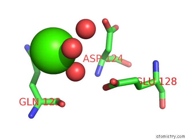
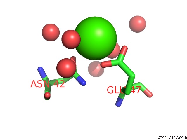
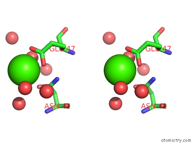
Calcium binding site 1 out of 4 in 5veq
Go back to
Calcium binding site 1 out
of 4 in the Mouse Kynurenine Aminotransferase III, Re-Refinement of the Pdb Structure 3E2Y
Mono view
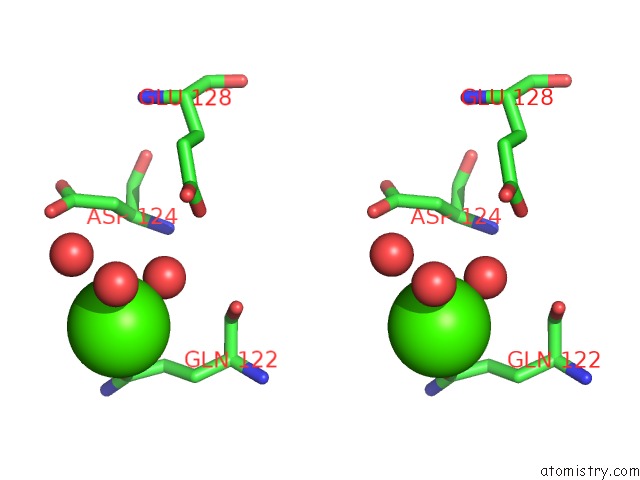
Stereo pair view
Mono view
Stereo pair view
A full contact list of Calcium with other atoms in the Ca binding
site number 1 of Mouse Kynurenine Aminotransferase III, Re-Refinement of the Pdb Structure 3E2Y within 5.0Å range:
|
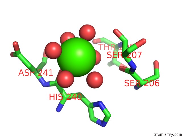
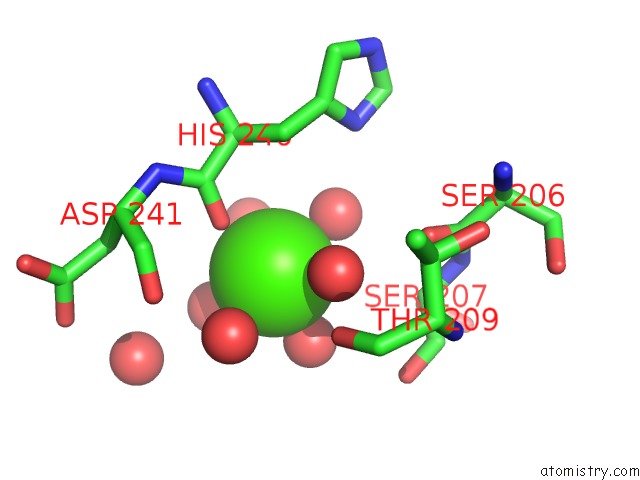
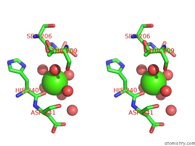
Calcium binding site 2 out of 4 in 5veq
Go back to
Calcium binding site 2 out
of 4 in the Mouse Kynurenine Aminotransferase III, Re-Refinement of the Pdb Structure 3E2Y
Mono view
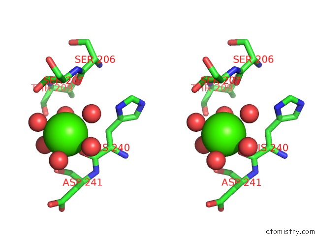
Stereo pair view
Mono view
Stereo pair view
A full contact list of Calcium with other atoms in the Ca binding
site number 2 of Mouse Kynurenine Aminotransferase III, Re-Refinement of the Pdb Structure 3E2Y within 5.0Å range:
|
Calcium binding site 3 out of 4 in 5veq
Go back to
Calcium binding site 3 out
of 4 in the Mouse Kynurenine Aminotransferase III, Re-Refinement of the Pdb Structure 3E2Y
Mono view
Stereo pair view
Mono view
Stereo pair view
A full contact list of Calcium with other atoms in the Ca binding
site number 3 of Mouse Kynurenine Aminotransferase III, Re-Refinement of the Pdb Structure 3E2Y within 5.0Å range:
|
Calcium binding site 4 out of 4 in 5veq
Go back to
Calcium binding site 4 out
of 4 in the Mouse Kynurenine Aminotransferase III, Re-Refinement of the Pdb Structure 3E2Y
Mono view
Stereo pair view
Mono view
Stereo pair view
A full contact list of Calcium with other atoms in the Ca binding
site number 4 of Mouse Kynurenine Aminotransferase III, Re-Refinement of the Pdb Structure 3E2Y within 5.0Å range:
|
Reference:
A.Wlodawer,
Z.Dauter,
P.J.Porebski,
W.Minor,
R.Stanfield,
M.Jaskolski,
E.Pozharski,
C.X.Weichenberger,
B.Rupp.
Detect, Correct, Retract: How to Manage Incorrect Structural Models. Febs J. V. 285 444 2018.
ISSN: ISSN 1742-4658
PubMed: 29113027
DOI: 10.1111/FEBS.14320
Page generated: Wed Jul 9 10:45:45 2025
ISSN: ISSN 1742-4658
PubMed: 29113027
DOI: 10.1111/FEBS.14320
Last articles
Mg in 4B3RMg in 4B9V
Mg in 4B9U
Mg in 4B9T
Mg in 4B9S
Mg in 4B9Q
Mg in 4B9N
Mg in 4B9M
Mg in 4B9L
Mg in 4B61