Calcium »
PDB 5vll-5w78 »
5vug »
Calcium in PDB 5vug: Crystal Structure of Glycerophosphoryl Diester Phosphodiesterase Domain of Uncharacterized Protein RV2277C From Mycobacterium Tuberculosis
Protein crystallography data
The structure of Crystal Structure of Glycerophosphoryl Diester Phosphodiesterase Domain of Uncharacterized Protein RV2277C From Mycobacterium Tuberculosis, PDB code: 5vug
was solved by
Y.Kim,
H.Li,
M.Endres,
A.Joachimiak,
Midwest Center For Structuralgenomics (Mcsg),
with X-Ray Crystallography technique. A brief refinement statistics is given in the table below:
| Resolution Low / High (Å) | 26.28 / 1.50 |
| Space group | P 21 21 2 |
| Cell size a, b, c (Å), α, β, γ (°) | 112.269, 43.878, 68.611, 90.00, 90.00, 90.00 |
| R / Rfree (%) | 16.3 / 19.6 |
Calcium Binding Sites:
The binding sites of Calcium atom in the Crystal Structure of Glycerophosphoryl Diester Phosphodiesterase Domain of Uncharacterized Protein RV2277C From Mycobacterium Tuberculosis
(pdb code 5vug). This binding sites where shown within
5.0 Angstroms radius around Calcium atom.
In total only one binding site of Calcium was determined in the Crystal Structure of Glycerophosphoryl Diester Phosphodiesterase Domain of Uncharacterized Protein RV2277C From Mycobacterium Tuberculosis, PDB code: 5vug:
In total only one binding site of Calcium was determined in the Crystal Structure of Glycerophosphoryl Diester Phosphodiesterase Domain of Uncharacterized Protein RV2277C From Mycobacterium Tuberculosis, PDB code: 5vug:
Calcium binding site 1 out of 1 in 5vug
Go back to
Calcium binding site 1 out
of 1 in the Crystal Structure of Glycerophosphoryl Diester Phosphodiesterase Domain of Uncharacterized Protein RV2277C From Mycobacterium Tuberculosis
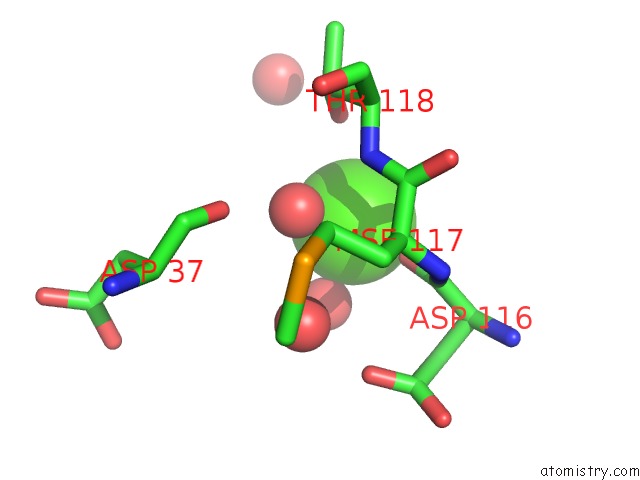
Mono view
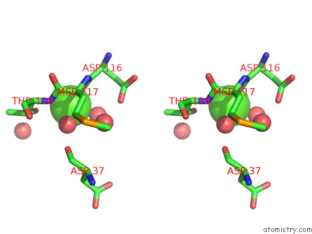
Stereo pair view
Mono view
Stereo pair view
A full contact list of Calcium with other atoms in the Ca binding
site number 1 of Crystal Structure of Glycerophosphoryl Diester Phosphodiesterase Domain of Uncharacterized Protein RV2277C From Mycobacterium Tuberculosis within 5.0Å range:
|
Reference:
Y.Kim,
H.Li,
M.Endres,
A.Joachimiak.
Crystal Structure of Glycerophosphoryl Diester Phosphodiesterase Domain of Uncharacterized Protein RV2277C From Mycobacterium Tuberculosis To Be Published.
Page generated: Wed Jul 9 10:53:53 2025
Last articles
Mg in 4DUXMg in 4DUW
Mg in 4DUV
Mg in 4DUO
Mg in 4DUG
Mg in 4DTY
Mg in 4DTW
Mg in 4DTH
Mg in 4DTF
Mg in 4DSU