Calcium »
PDB 5w7a-5wn3 »
5wey »
Calcium in PDB 5wey: Joint X-Ray/Neutron Structure of Concanavalin A with ALPHA1-2 D- Mannobiose
Protein crystallography data
The structure of Joint X-Ray/Neutron Structure of Concanavalin A with ALPHA1-2 D- Mannobiose, PDB code: 5wey
was solved by
A.Kovalevsky,
O.O.Gerlits,
R.J.Woods,
with X-Ray Crystallography technique. A brief refinement statistics is given in the table below:
| Resolution Low / High (Å) | N/A / 1.80 |
| Space group | I 2 2 2 |
| Cell size a, b, c (Å), α, β, γ (°) | 66.563, 86.618, 91.808, 90.00, 90.00, 90.00 |
| R / Rfree (%) | 24.7 / 28.5 |
Other elements in 5wey:
The structure of Joint X-Ray/Neutron Structure of Concanavalin A with ALPHA1-2 D- Mannobiose also contains other interesting chemical elements:
| Manganese | (Mn) | 1 atom |
Calcium Binding Sites:
The binding sites of Calcium atom in the Joint X-Ray/Neutron Structure of Concanavalin A with ALPHA1-2 D- Mannobiose
(pdb code 5wey). This binding sites where shown within
5.0 Angstroms radius around Calcium atom.
In total only one binding site of Calcium was determined in the Joint X-Ray/Neutron Structure of Concanavalin A with ALPHA1-2 D- Mannobiose, PDB code: 5wey:
In total only one binding site of Calcium was determined in the Joint X-Ray/Neutron Structure of Concanavalin A with ALPHA1-2 D- Mannobiose, PDB code: 5wey:
Calcium binding site 1 out of 1 in 5wey
Go back to
Calcium binding site 1 out
of 1 in the Joint X-Ray/Neutron Structure of Concanavalin A with ALPHA1-2 D- Mannobiose
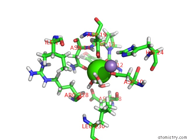
Mono view
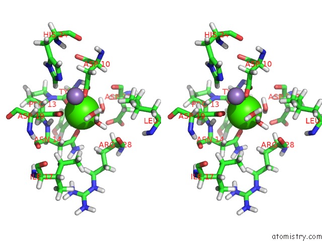
Stereo pair view
Mono view
Stereo pair view
A full contact list of Calcium with other atoms in the Ca binding
site number 1 of Joint X-Ray/Neutron Structure of Concanavalin A with ALPHA1-2 D- Mannobiose within 5.0Å range:
|
Reference:
O.O.Gerlits,
L.Coates,
R.J.Woods,
A.Kovalevsky.
Mannobiose Binding Induces Changes in Hydrogen Bonding and Protonation States of Acidic Residues in Concanavalin A As Revealed By Neutron Crystallography. Biochemistry V. 56 4747 2017.
ISSN: ISSN 1520-4995
PubMed: 28846383
DOI: 10.1021/ACS.BIOCHEM.7B00654
Page generated: Wed Jul 9 11:12:18 2025
ISSN: ISSN 1520-4995
PubMed: 28846383
DOI: 10.1021/ACS.BIOCHEM.7B00654
Last articles
Mg in 5SC9Mg in 5SCB
Mg in 5SC8
Mg in 5SCA
Mg in 5SBE
Mg in 5SBD
Mg in 5SBC
Mg in 5SBB
Mg in 5SBA
Mg in 5SB8