Calcium »
PDB 5w7a-5wn3 »
5wkj »
Calcium in PDB 5wkj: 2.05 A Resolution Structure of Mers 3CL Protease in Complex with Inhibitor GC376
Protein crystallography data
The structure of 2.05 A Resolution Structure of Mers 3CL Protease in Complex with Inhibitor GC376, PDB code: 5wkj
was solved by
S.Lovell,
K.P.Battaile,
N.Mehzabeen,
A.C.G.Kankanamalage,
Y.Kim,
A.D.Rathnayake,
K.O.Chang,
W.C.Groutas,
with X-Ray Crystallography technique. A brief refinement statistics is given in the table below:
| Resolution Low / High (Å) | 41.79 / 2.05 |
| Space group | C 1 2 1 |
| Cell size a, b, c (Å), α, β, γ (°) | 101.807, 58.292, 49.785, 90.00, 112.25, 90.00 |
| R / Rfree (%) | 16 / 23.4 |
Calcium Binding Sites:
The binding sites of Calcium atom in the 2.05 A Resolution Structure of Mers 3CL Protease in Complex with Inhibitor GC376
(pdb code 5wkj). This binding sites where shown within
5.0 Angstroms radius around Calcium atom.
In total 2 binding sites of Calcium where determined in the 2.05 A Resolution Structure of Mers 3CL Protease in Complex with Inhibitor GC376, PDB code: 5wkj:
Jump to Calcium binding site number: 1; 2;
In total 2 binding sites of Calcium where determined in the 2.05 A Resolution Structure of Mers 3CL Protease in Complex with Inhibitor GC376, PDB code: 5wkj:
Jump to Calcium binding site number: 1; 2;
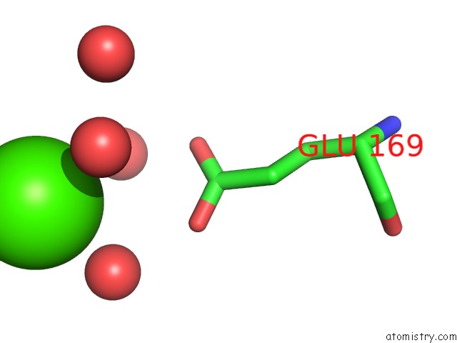
Calcium binding site 1 out of 2 in 5wkj
Go back to
Calcium binding site 1 out
of 2 in the 2.05 A Resolution Structure of Mers 3CL Protease in Complex with Inhibitor GC376
Mono view
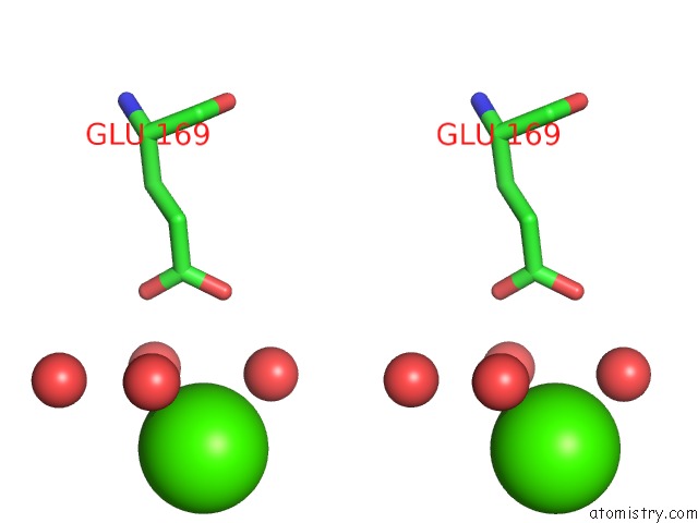
Stereo pair view
Mono view
Stereo pair view
A full contact list of Calcium with other atoms in the Ca binding
site number 1 of 2.05 A Resolution Structure of Mers 3CL Protease in Complex with Inhibitor GC376 within 5.0Å range:
|
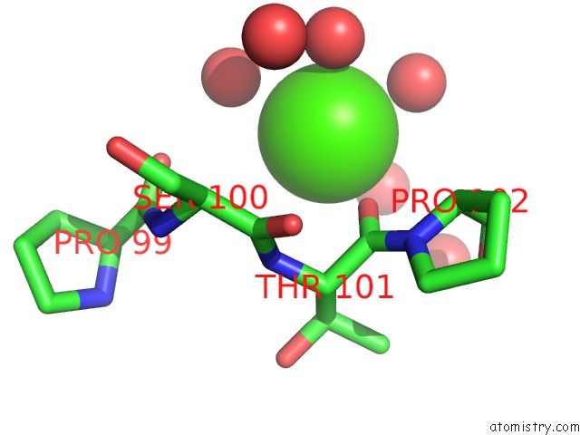
Calcium binding site 2 out of 2 in 5wkj
Go back to
Calcium binding site 2 out
of 2 in the 2.05 A Resolution Structure of Mers 3CL Protease in Complex with Inhibitor GC376
Mono view
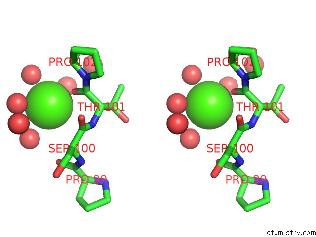
Stereo pair view
Mono view
Stereo pair view
A full contact list of Calcium with other atoms in the Ca binding
site number 2 of 2.05 A Resolution Structure of Mers 3CL Protease in Complex with Inhibitor GC376 within 5.0Å range:
|
Reference:
A.C.Galasiti Kankanamalage,
Y.Kim,
V.C.Damalanka,
A.D.Rathnayake,
A.R.Fehr,
N.Mehzabeen,
K.P.Battaile,
S.Lovell,
G.H.Lushington,
S.Perlman,
K.O.Chang,
W.C.Groutas.
Structure-Guided Design of Potent and Permeable Inhibitors of Mers Coronavirus 3CL Protease That Utilize A Piperidine Moiety As A Novel Design Element. Eur J Med Chem V. 150 334 2018.
ISSN: ISSN 1768-3254
PubMed: 29544147
DOI: 10.1016/J.EJMECH.2018.03.004
Page generated: Wed Jul 9 11:17:00 2025
ISSN: ISSN 1768-3254
PubMed: 29544147
DOI: 10.1016/J.EJMECH.2018.03.004
Last articles
Mg in 4DPGMg in 4DQP
Mg in 4DQQ
Mg in 4DPM
Mg in 4DPV
Mg in 4DQI
Mg in 4DOB
Mg in 4DOC
Mg in 4DMZ
Mg in 4DOA