Calcium »
PDB 5w7a-5wn3 »
5wmm »
Calcium in PDB 5wmm: Crystal Structure of An Adenylation Domain Interrupted By A Methylation Domain (AMA4) From Nonribosomal Peptide Synthetase Tios
Protein crystallography data
The structure of Crystal Structure of An Adenylation Domain Interrupted By A Methylation Domain (AMA4) From Nonribosomal Peptide Synthetase Tios, PDB code: 5wmm
was solved by
A.H.Pang,
S.Mori,
S.Garneau-Tsodikova,
O.V.Tsodikov,
with X-Ray Crystallography technique. A brief refinement statistics is given in the table below:
| Resolution Low / High (Å) | 40.00 / 2.90 |
| Space group | P 32 2 1 |
| Cell size a, b, c (Å), α, β, γ (°) | 136.712, 136.712, 228.234, 90.00, 90.00, 120.00 |
| R / Rfree (%) | 23.6 / 25.3 |
Other elements in 5wmm:
The structure of Crystal Structure of An Adenylation Domain Interrupted By A Methylation Domain (AMA4) From Nonribosomal Peptide Synthetase Tios also contains other interesting chemical elements:
| Chlorine | (Cl) | 3 atoms |
Calcium Binding Sites:
The binding sites of Calcium atom in the Crystal Structure of An Adenylation Domain Interrupted By A Methylation Domain (AMA4) From Nonribosomal Peptide Synthetase Tios
(pdb code 5wmm). This binding sites where shown within
5.0 Angstroms radius around Calcium atom.
In total 3 binding sites of Calcium where determined in the Crystal Structure of An Adenylation Domain Interrupted By A Methylation Domain (AMA4) From Nonribosomal Peptide Synthetase Tios, PDB code: 5wmm:
Jump to Calcium binding site number: 1; 2; 3;
In total 3 binding sites of Calcium where determined in the Crystal Structure of An Adenylation Domain Interrupted By A Methylation Domain (AMA4) From Nonribosomal Peptide Synthetase Tios, PDB code: 5wmm:
Jump to Calcium binding site number: 1; 2; 3;
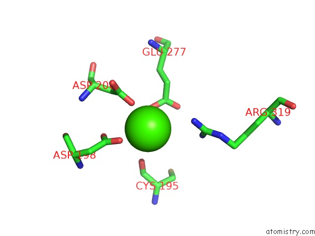
Calcium binding site 1 out of 3 in 5wmm
Go back to
Calcium binding site 1 out
of 3 in the Crystal Structure of An Adenylation Domain Interrupted By A Methylation Domain (AMA4) From Nonribosomal Peptide Synthetase Tios
Mono view
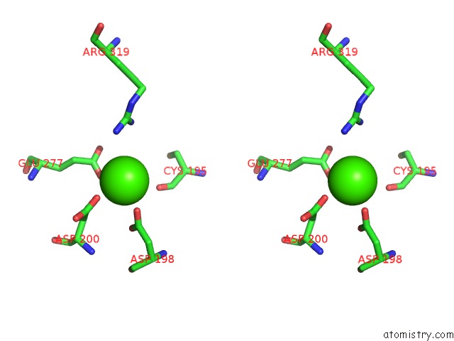
Stereo pair view
Mono view
Stereo pair view
A full contact list of Calcium with other atoms in the Ca binding
site number 1 of Crystal Structure of An Adenylation Domain Interrupted By A Methylation Domain (AMA4) From Nonribosomal Peptide Synthetase Tios within 5.0Å range:
|
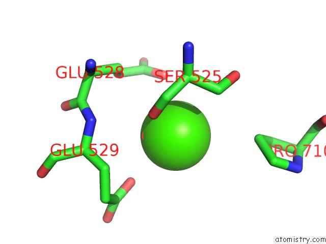
Calcium binding site 2 out of 3 in 5wmm
Go back to
Calcium binding site 2 out
of 3 in the Crystal Structure of An Adenylation Domain Interrupted By A Methylation Domain (AMA4) From Nonribosomal Peptide Synthetase Tios
Mono view
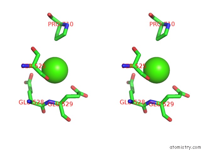
Stereo pair view
Mono view
Stereo pair view
A full contact list of Calcium with other atoms in the Ca binding
site number 2 of Crystal Structure of An Adenylation Domain Interrupted By A Methylation Domain (AMA4) From Nonribosomal Peptide Synthetase Tios within 5.0Å range:
|
Calcium binding site 3 out of 3 in 5wmm
Go back to
Calcium binding site 3 out
of 3 in the Crystal Structure of An Adenylation Domain Interrupted By A Methylation Domain (AMA4) From Nonribosomal Peptide Synthetase Tios

Mono view

Stereo pair view
Mono view
Stereo pair view
A full contact list of Calcium with other atoms in the Ca binding
site number 3 of Crystal Structure of An Adenylation Domain Interrupted By A Methylation Domain (AMA4) From Nonribosomal Peptide Synthetase Tios within 5.0Å range:
|
Reference:
S.Mori,
A.H.Pang,
T.A.Lundy,
A.Garzan,
O.V.Tsodikov,
S.Garneau-Tsodikova.
Structural Basis For Backbone N-Methylation By An Interrupted Adenylation Domain. Nat. Chem. Biol. V. 14 428 2018.
ISSN: ESSN 1552-4469
PubMed: 29556104
DOI: 10.1038/S41589-018-0014-7
Page generated: Wed Jul 9 11:17:27 2025
ISSN: ESSN 1552-4469
PubMed: 29556104
DOI: 10.1038/S41589-018-0014-7
Last articles
Mg in 3SE1Mg in 3SE5
Mg in 3SDK
Mg in 3SDV
Mg in 3SDU
Mg in 3SDT
Mg in 3SDR
Mg in 3SBF
Mg in 3SB5
Mg in 3SCY