Calcium »
PDB 5xkf-5xu9 »
5xts »
Calcium in PDB 5xts: Crystal Structure of the Cysr-CTLD3 Fragment of Human Mr at Basic/Neutral pH
Protein crystallography data
The structure of Crystal Structure of the Cysr-CTLD3 Fragment of Human Mr at Basic/Neutral pH, PDB code: 5xts
was solved by
Y.He,
Z.Hu,
with X-Ray Crystallography technique. A brief refinement statistics is given in the table below:
| Resolution Low / High (Å) | 38.86 / 2.00 |
| Space group | P 21 21 21 |
| Cell size a, b, c (Å), α, β, γ (°) | 60.256, 101.697, 123.464, 90.00, 90.00, 90.00 |
| R / Rfree (%) | 16.6 / 20.1 |
Calcium Binding Sites:
The binding sites of Calcium atom in the Crystal Structure of the Cysr-CTLD3 Fragment of Human Mr at Basic/Neutral pH
(pdb code 5xts). This binding sites where shown within
5.0 Angstroms radius around Calcium atom.
In total only one binding site of Calcium was determined in the Crystal Structure of the Cysr-CTLD3 Fragment of Human Mr at Basic/Neutral pH, PDB code: 5xts:
In total only one binding site of Calcium was determined in the Crystal Structure of the Cysr-CTLD3 Fragment of Human Mr at Basic/Neutral pH, PDB code: 5xts:
Calcium binding site 1 out of 1 in 5xts
Go back to
Calcium binding site 1 out
of 1 in the Crystal Structure of the Cysr-CTLD3 Fragment of Human Mr at Basic/Neutral pH
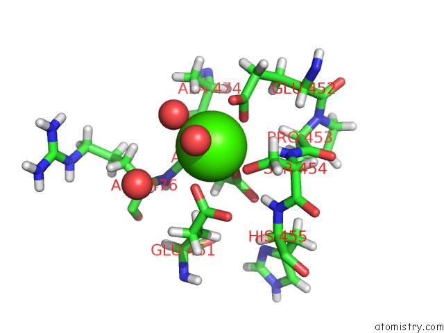
Mono view
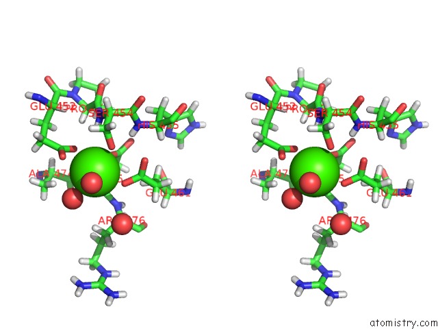
Stereo pair view
Mono view
Stereo pair view
A full contact list of Calcium with other atoms in the Ca binding
site number 1 of Crystal Structure of the Cysr-CTLD3 Fragment of Human Mr at Basic/Neutral pH within 5.0Å range:
|
Reference:
Z.Hu,
X.Shi,
B.Yu,
N.Li,
Y.Huang,
Y.He.
Structural Insights Into the pH-Dependent Conformational Change and Collagen Recognition of the Human Mannose Receptor Structure V. 26 60 2018.
ISSN: ISSN 1878-4186
PubMed: 29225077
DOI: 10.1016/J.STR.2017.11.006
Page generated: Wed Jul 9 11:47:19 2025
ISSN: ISSN 1878-4186
PubMed: 29225077
DOI: 10.1016/J.STR.2017.11.006
Last articles
Mg in 4DV3Mg in 4DV2
Mg in 4DV0
Mg in 4DV1
Mg in 4DUZ
Mg in 4DUY
Mg in 4DR7
Mg in 4DR6
Mg in 4DR5
Mg in 4DUX