Calcium »
PDB 5yjd-5z4p »
5ynd »
Calcium in PDB 5ynd: Crystal Structure of Pullulanase From Klebsiella Pneumoniae Complex at 1 Mm Gamma-Cyclodextrin
Protein crystallography data
The structure of Crystal Structure of Pullulanase From Klebsiella Pneumoniae Complex at 1 Mm Gamma-Cyclodextrin, PDB code: 5ynd
was solved by
N.Saka,
H.Iwamoto,
N.Takahashi,
K.Mizutani,
B.Mikami,
with X-Ray Crystallography technique. A brief refinement statistics is given in the table below:
| Resolution Low / High (Å) | 44.39 / 2.23 |
| Space group | P 43 21 2 |
| Cell size a, b, c (Å), α, β, γ (°) | 88.773, 88.773, 298.379, 90.00, 90.00, 90.00 |
| R / Rfree (%) | 19.1 / 24.3 |
Calcium Binding Sites:
The binding sites of Calcium atom in the Crystal Structure of Pullulanase From Klebsiella Pneumoniae Complex at 1 Mm Gamma-Cyclodextrin
(pdb code 5ynd). This binding sites where shown within
5.0 Angstroms radius around Calcium atom.
In total 5 binding sites of Calcium where determined in the Crystal Structure of Pullulanase From Klebsiella Pneumoniae Complex at 1 Mm Gamma-Cyclodextrin, PDB code: 5ynd:
Jump to Calcium binding site number: 1; 2; 3; 4; 5;
In total 5 binding sites of Calcium where determined in the Crystal Structure of Pullulanase From Klebsiella Pneumoniae Complex at 1 Mm Gamma-Cyclodextrin, PDB code: 5ynd:
Jump to Calcium binding site number: 1; 2; 3; 4; 5;
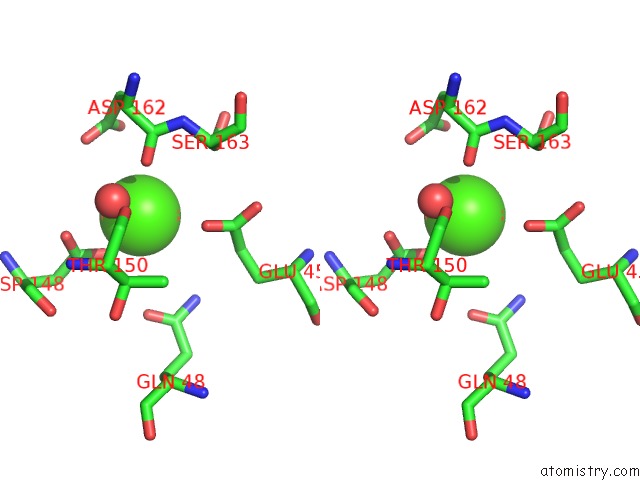
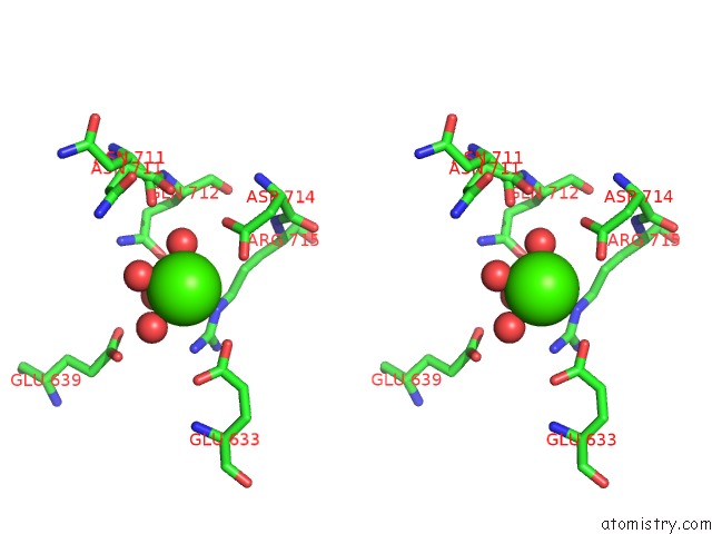
Calcium binding site 1 out of 5 in 5ynd
Go back to
Calcium binding site 1 out
of 5 in the Crystal Structure of Pullulanase From Klebsiella Pneumoniae Complex at 1 Mm Gamma-Cyclodextrin
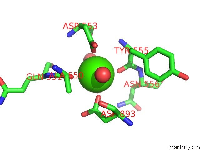
Mono view

Stereo pair view
Mono view
Stereo pair view
A full contact list of Calcium with other atoms in the Ca binding
site number 1 of Crystal Structure of Pullulanase From Klebsiella Pneumoniae Complex at 1 Mm Gamma-Cyclodextrin within 5.0Å range:
|
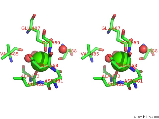
Calcium binding site 2 out of 5 in 5ynd
Go back to
Calcium binding site 2 out
of 5 in the Crystal Structure of Pullulanase From Klebsiella Pneumoniae Complex at 1 Mm Gamma-Cyclodextrin
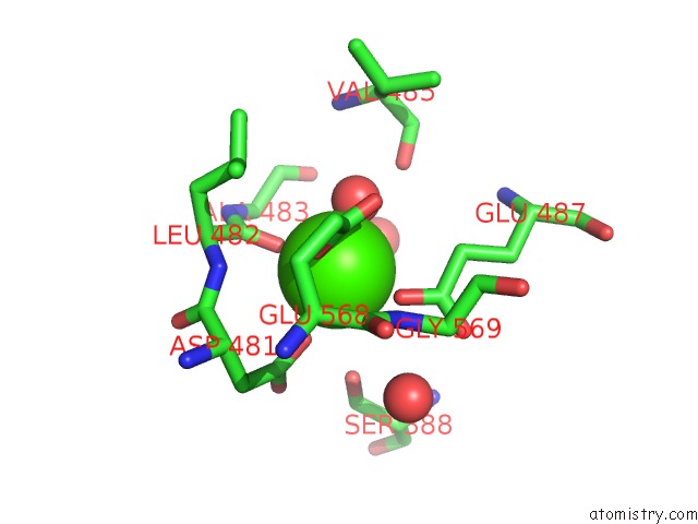
Mono view
Stereo pair view
Mono view
Stereo pair view
A full contact list of Calcium with other atoms in the Ca binding
site number 2 of Crystal Structure of Pullulanase From Klebsiella Pneumoniae Complex at 1 Mm Gamma-Cyclodextrin within 5.0Å range:
|
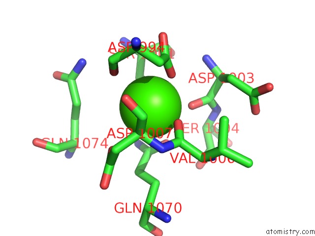
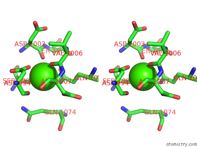
Calcium binding site 3 out of 5 in 5ynd
Go back to
Calcium binding site 3 out
of 5 in the Crystal Structure of Pullulanase From Klebsiella Pneumoniae Complex at 1 Mm Gamma-Cyclodextrin
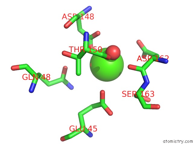
Mono view
Stereo pair view
Mono view
Stereo pair view
A full contact list of Calcium with other atoms in the Ca binding
site number 3 of Crystal Structure of Pullulanase From Klebsiella Pneumoniae Complex at 1 Mm Gamma-Cyclodextrin within 5.0Å range:
|
Calcium binding site 4 out of 5 in 5ynd
Go back to
Calcium binding site 4 out
of 5 in the Crystal Structure of Pullulanase From Klebsiella Pneumoniae Complex at 1 Mm Gamma-Cyclodextrin
Mono view
Stereo pair view
Mono view
Stereo pair view
A full contact list of Calcium with other atoms in the Ca binding
site number 4 of Crystal Structure of Pullulanase From Klebsiella Pneumoniae Complex at 1 Mm Gamma-Cyclodextrin within 5.0Å range:
|
Calcium binding site 5 out of 5 in 5ynd
Go back to
Calcium binding site 5 out
of 5 in the Crystal Structure of Pullulanase From Klebsiella Pneumoniae Complex at 1 Mm Gamma-Cyclodextrin
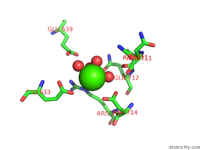
Mono view
Stereo pair view
Mono view
Stereo pair view
A full contact list of Calcium with other atoms in the Ca binding
site number 5 of Crystal Structure of Pullulanase From Klebsiella Pneumoniae Complex at 1 Mm Gamma-Cyclodextrin within 5.0Å range:
|
Reference:
N.Saka,
H.Iwamoto,
D.Malle,
N.Takahashi,
K.Mizutani,
B.Mikami.
Elucidation of the Mechanism of Interaction Between Klebsiella Pneumoniae Pullulanase and Cyclodextrin Acta Crystallogr D Struct V. 74 1115 2018BIOL.
ISSN: ISSN 2059-7983
PubMed: 30387770
DOI: 10.1107/S2059798318014523
Page generated: Wed Jul 9 12:01:12 2025
ISSN: ISSN 2059-7983
PubMed: 30387770
DOI: 10.1107/S2059798318014523
Last articles
K in 3FWFK in 3FWG
K in 3FWJ
K in 3FWI
K in 3FTM
K in 3FTF
K in 3FPB
K in 3FN2
K in 3FKW
K in 3FKV