Calcium »
PDB 5yjd-5z4p »
5ypu »
Calcium in PDB 5ypu: Crystal Structure of An Actin Monomer in Complex with the Nucleator Cordon-Bleu MET72NLE WH2-Motif Peptide
Protein crystallography data
The structure of Crystal Structure of An Actin Monomer in Complex with the Nucleator Cordon-Bleu MET72NLE WH2-Motif Peptide, PDB code: 5ypu
was solved by
C.P.M.Scipion,
J.Wongsantichon,
F.J.Ferrer,
T.Y.Yuen,
R.C.Robinson,
with X-Ray Crystallography technique. A brief refinement statistics is given in the table below:
| Resolution Low / High (Å) | 29.82 / 2.00 |
| Space group | C 1 2 1 |
| Cell size a, b, c (Å), α, β, γ (°) | 174.861, 40.832, 109.000, 90.00, 101.76, 90.00 |
| R / Rfree (%) | 19.3 / 24 |
Calcium Binding Sites:
The binding sites of Calcium atom in the Crystal Structure of An Actin Monomer in Complex with the Nucleator Cordon-Bleu MET72NLE WH2-Motif Peptide
(pdb code 5ypu). This binding sites where shown within
5.0 Angstroms radius around Calcium atom.
In total 7 binding sites of Calcium where determined in the Crystal Structure of An Actin Monomer in Complex with the Nucleator Cordon-Bleu MET72NLE WH2-Motif Peptide, PDB code: 5ypu:
Jump to Calcium binding site number: 1; 2; 3; 4; 5; 6; 7;
In total 7 binding sites of Calcium where determined in the Crystal Structure of An Actin Monomer in Complex with the Nucleator Cordon-Bleu MET72NLE WH2-Motif Peptide, PDB code: 5ypu:
Jump to Calcium binding site number: 1; 2; 3; 4; 5; 6; 7;
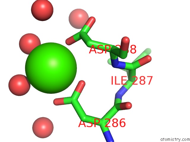
Calcium binding site 1 out of 7 in 5ypu
Go back to
Calcium binding site 1 out
of 7 in the Crystal Structure of An Actin Monomer in Complex with the Nucleator Cordon-Bleu MET72NLE WH2-Motif Peptide

Mono view

Stereo pair view
Mono view
Stereo pair view
A full contact list of Calcium with other atoms in the Ca binding
site number 1 of Crystal Structure of An Actin Monomer in Complex with the Nucleator Cordon-Bleu MET72NLE WH2-Motif Peptide within 5.0Å range:
|
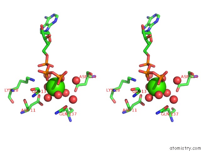
Calcium binding site 2 out of 7 in 5ypu
Go back to
Calcium binding site 2 out
of 7 in the Crystal Structure of An Actin Monomer in Complex with the Nucleator Cordon-Bleu MET72NLE WH2-Motif Peptide
Mono view

Stereo pair view
Mono view
Stereo pair view
A full contact list of Calcium with other atoms in the Ca binding
site number 2 of Crystal Structure of An Actin Monomer in Complex with the Nucleator Cordon-Bleu MET72NLE WH2-Motif Peptide within 5.0Å range:
|
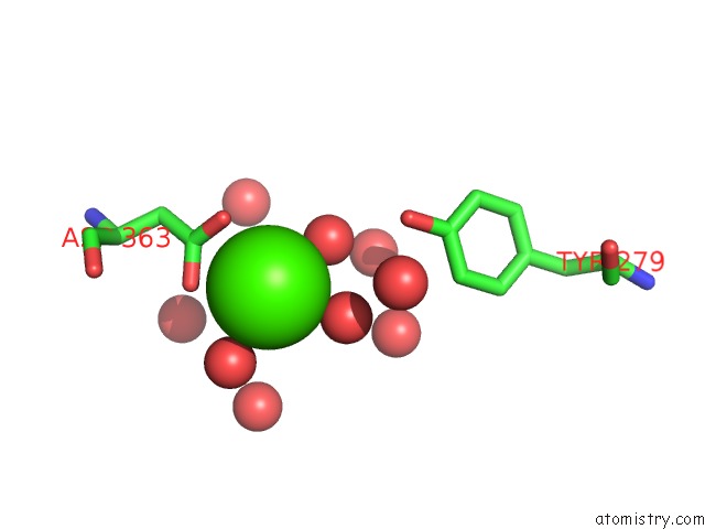
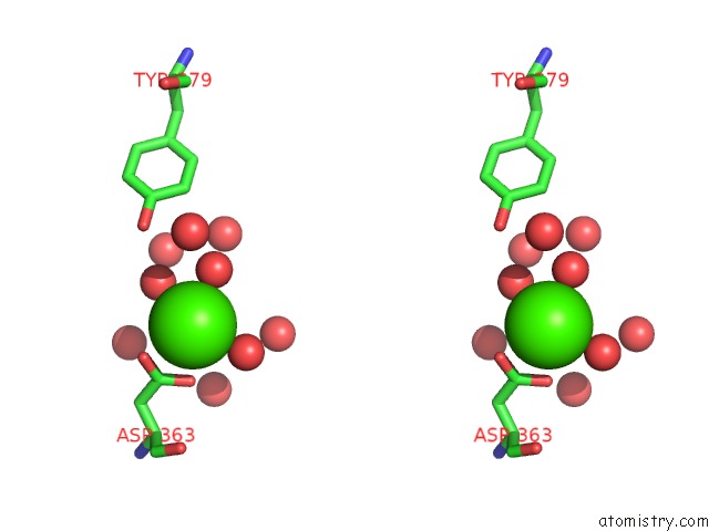
Calcium binding site 3 out of 7 in 5ypu
Go back to
Calcium binding site 3 out
of 7 in the Crystal Structure of An Actin Monomer in Complex with the Nucleator Cordon-Bleu MET72NLE WH2-Motif Peptide

Mono view

Stereo pair view
Mono view
Stereo pair view
A full contact list of Calcium with other atoms in the Ca binding
site number 3 of Crystal Structure of An Actin Monomer in Complex with the Nucleator Cordon-Bleu MET72NLE WH2-Motif Peptide within 5.0Å range:
|
Calcium binding site 4 out of 7 in 5ypu
Go back to
Calcium binding site 4 out
of 7 in the Crystal Structure of An Actin Monomer in Complex with the Nucleator Cordon-Bleu MET72NLE WH2-Motif Peptide

Mono view
Stereo pair view
Mono view
Stereo pair view
A full contact list of Calcium with other atoms in the Ca binding
site number 4 of Crystal Structure of An Actin Monomer in Complex with the Nucleator Cordon-Bleu MET72NLE WH2-Motif Peptide within 5.0Å range:
|
Calcium binding site 5 out of 7 in 5ypu
Go back to
Calcium binding site 5 out
of 7 in the Crystal Structure of An Actin Monomer in Complex with the Nucleator Cordon-Bleu MET72NLE WH2-Motif Peptide

Mono view

Stereo pair view
Mono view
Stereo pair view
A full contact list of Calcium with other atoms in the Ca binding
site number 5 of Crystal Structure of An Actin Monomer in Complex with the Nucleator Cordon-Bleu MET72NLE WH2-Motif Peptide within 5.0Å range:
|
Calcium binding site 6 out of 7 in 5ypu
Go back to
Calcium binding site 6 out
of 7 in the Crystal Structure of An Actin Monomer in Complex with the Nucleator Cordon-Bleu MET72NLE WH2-Motif Peptide
Mono view
Stereo pair view
Mono view
Stereo pair view
A full contact list of Calcium with other atoms in the Ca binding
site number 6 of Crystal Structure of An Actin Monomer in Complex with the Nucleator Cordon-Bleu MET72NLE WH2-Motif Peptide within 5.0Å range:
|
Calcium binding site 7 out of 7 in 5ypu
Go back to
Calcium binding site 7 out
of 7 in the Crystal Structure of An Actin Monomer in Complex with the Nucleator Cordon-Bleu MET72NLE WH2-Motif Peptide

Mono view

Stereo pair view
Mono view
Stereo pair view
A full contact list of Calcium with other atoms in the Ca binding
site number 7 of Crystal Structure of An Actin Monomer in Complex with the Nucleator Cordon-Bleu MET72NLE WH2-Motif Peptide within 5.0Å range:
|
Reference:
C.P.M.Scipion,
U.Ghoshdastider,
F.J.Ferrer,
T.Y.Yuen,
J.Wongsantichon,
R.C.Robinson.
Structural Evidence For the Roles of Divalent Cations in Actin Polymerization and Activation of Atp Hydrolysis Proc. Natl. Acad. Sci. V. 115 10345 2018U.S.A..
ISSN: ESSN 1091-6490
PubMed: 30254171
DOI: 10.1073/PNAS.1806394115
Page generated: Wed Jul 9 12:02:43 2025
ISSN: ESSN 1091-6490
PubMed: 30254171
DOI: 10.1073/PNAS.1806394115
Last articles
K in 3HJWK in 3HFW
K in 3HC1
K in 3GVX
K in 3H7C
K in 3GX3
K in 3H4K
K in 3GX5
K in 3G78
K in 3GKO