Calcium »
PDB 5yjd-5z4p »
5ysh »
Calcium in PDB 5ysh: Diol Dehydratase - Alpha/T172A Mutant Complexed with Adocbl, Aerobically-Prepared Crystal
Enzymatic activity of Diol Dehydratase - Alpha/T172A Mutant Complexed with Adocbl, Aerobically-Prepared Crystal
All present enzymatic activity of Diol Dehydratase - Alpha/T172A Mutant Complexed with Adocbl, Aerobically-Prepared Crystal:
4.2.1.28;
4.2.1.28;
Protein crystallography data
The structure of Diol Dehydratase - Alpha/T172A Mutant Complexed with Adocbl, Aerobically-Prepared Crystal, PDB code: 5ysh
was solved by
N.Shibata,
with X-Ray Crystallography technique. A brief refinement statistics is given in the table below:
| Resolution Low / High (Å) | 49.00 / 1.90 |
| Space group | P 1 |
| Cell size a, b, c (Å), α, β, γ (°) | 70.330, 110.490, 115.480, 92.69, 95.97, 105.44 |
| R / Rfree (%) | 16.4 / 19.1 |
Other elements in 5ysh:
The structure of Diol Dehydratase - Alpha/T172A Mutant Complexed with Adocbl, Aerobically-Prepared Crystal also contains other interesting chemical elements:
| Cobalt | (Co) | 4 atoms |
| Potassium | (K) | 8 atoms |
Calcium Binding Sites:
The binding sites of Calcium atom in the Diol Dehydratase - Alpha/T172A Mutant Complexed with Adocbl, Aerobically-Prepared Crystal
(pdb code 5ysh). This binding sites where shown within
5.0 Angstroms radius around Calcium atom.
In total 4 binding sites of Calcium where determined in the Diol Dehydratase - Alpha/T172A Mutant Complexed with Adocbl, Aerobically-Prepared Crystal, PDB code: 5ysh:
Jump to Calcium binding site number: 1; 2; 3; 4;
In total 4 binding sites of Calcium where determined in the Diol Dehydratase - Alpha/T172A Mutant Complexed with Adocbl, Aerobically-Prepared Crystal, PDB code: 5ysh:
Jump to Calcium binding site number: 1; 2; 3; 4;
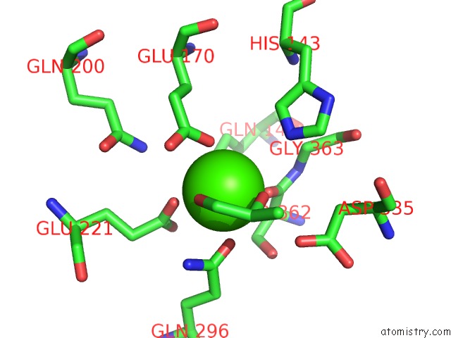
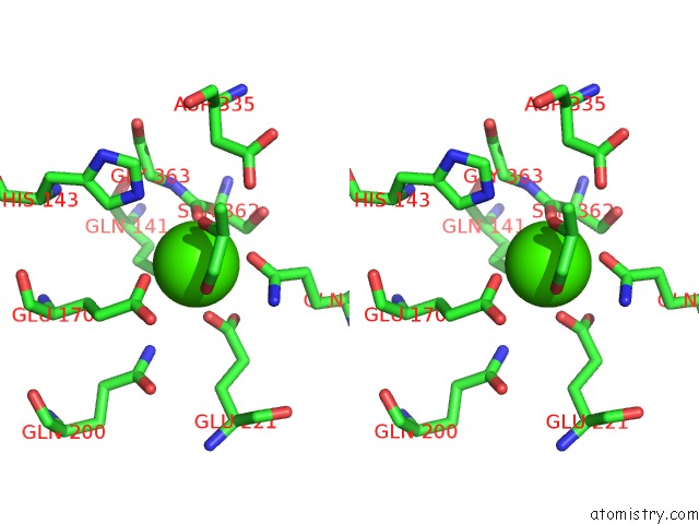
Calcium binding site 1 out of 4 in 5ysh
Go back to
Calcium binding site 1 out
of 4 in the Diol Dehydratase - Alpha/T172A Mutant Complexed with Adocbl, Aerobically-Prepared Crystal
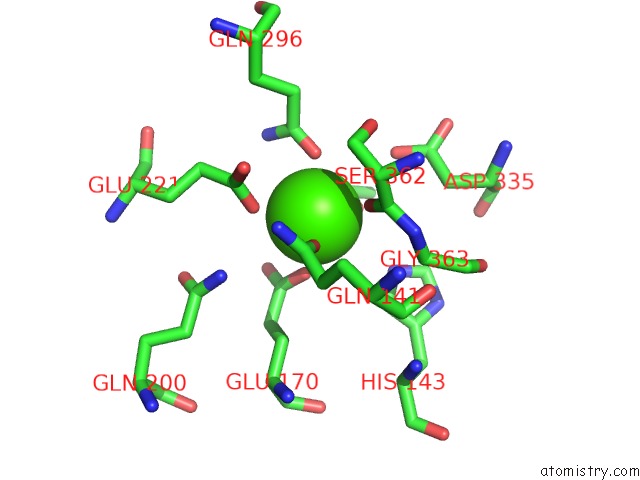
Mono view
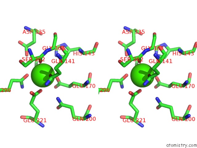
Stereo pair view
Mono view
Stereo pair view
A full contact list of Calcium with other atoms in the Ca binding
site number 1 of Diol Dehydratase - Alpha/T172A Mutant Complexed with Adocbl, Aerobically-Prepared Crystal within 5.0Å range:
|
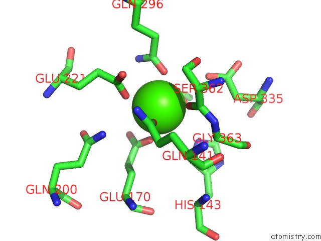
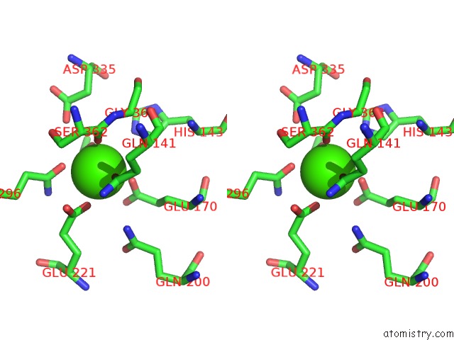
Calcium binding site 2 out of 4 in 5ysh
Go back to
Calcium binding site 2 out
of 4 in the Diol Dehydratase - Alpha/T172A Mutant Complexed with Adocbl, Aerobically-Prepared Crystal
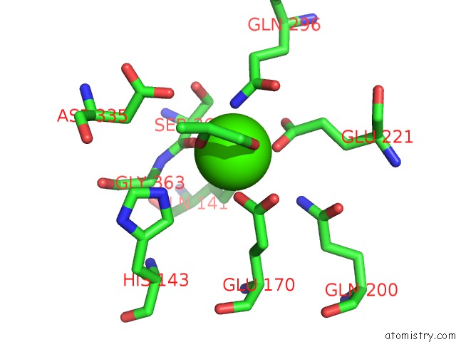
Mono view
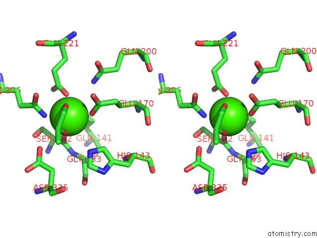
Stereo pair view
Mono view
Stereo pair view
A full contact list of Calcium with other atoms in the Ca binding
site number 2 of Diol Dehydratase - Alpha/T172A Mutant Complexed with Adocbl, Aerobically-Prepared Crystal within 5.0Å range:
|
Calcium binding site 3 out of 4 in 5ysh
Go back to
Calcium binding site 3 out
of 4 in the Diol Dehydratase - Alpha/T172A Mutant Complexed with Adocbl, Aerobically-Prepared Crystal
Mono view
Stereo pair view
Mono view
Stereo pair view
A full contact list of Calcium with other atoms in the Ca binding
site number 3 of Diol Dehydratase - Alpha/T172A Mutant Complexed with Adocbl, Aerobically-Prepared Crystal within 5.0Å range:
|
Calcium binding site 4 out of 4 in 5ysh
Go back to
Calcium binding site 4 out
of 4 in the Diol Dehydratase - Alpha/T172A Mutant Complexed with Adocbl, Aerobically-Prepared Crystal
Mono view
Stereo pair view
Mono view
Stereo pair view
A full contact list of Calcium with other atoms in the Ca binding
site number 4 of Diol Dehydratase - Alpha/T172A Mutant Complexed with Adocbl, Aerobically-Prepared Crystal within 5.0Å range:
|
Reference:
N.Shibata,
Y.Sueyoshi,
Y.Higuchi,
T.Toraya.
Direct Participation of A Peripheral Side Chain of A Corrin Ring in Coenzyme B12CATALYSIS. Angew. Chem. Int. Ed. Engl. V. 57 7830 2018.
ISSN: ESSN 1521-3773
PubMed: 29797764
DOI: 10.1002/ANIE.201803591
Page generated: Wed Jul 9 12:04:07 2025
ISSN: ESSN 1521-3773
PubMed: 29797764
DOI: 10.1002/ANIE.201803591
Last articles
Mg in 3CMQMg in 3CLY
Mg in 3CK5
Mg in 3CLC
Mg in 3CKG
Mg in 3CKE
Mg in 3CK4
Mg in 3CJO
Mg in 3CJ8
Mg in 3CIK