Calcium »
PDB 5yjd-5z4p »
5yua »
Calcium in PDB 5yua: Crystal Structure of Voltage-Gated Sodium Channel Navab in High-pH Condition
Protein crystallography data
The structure of Crystal Structure of Voltage-Gated Sodium Channel Navab in High-pH Condition, PDB code: 5yua
was solved by
K.Irie,
T.Shimomura,
Y.Fujiyoshi,
with X-Ray Crystallography technique. A brief refinement statistics is given in the table below:
| Resolution Low / High (Å) | 31.46 / 2.80 |
| Space group | I 4 2 2 |
| Cell size a, b, c (Å), α, β, γ (°) | 126.450, 126.450, 201.658, 90.00, 90.00, 90.00 |
| R / Rfree (%) | 23 / 26.2 |
Calcium Binding Sites:
The binding sites of Calcium atom in the Crystal Structure of Voltage-Gated Sodium Channel Navab in High-pH Condition
(pdb code 5yua). This binding sites where shown within
5.0 Angstroms radius around Calcium atom.
In total only one binding site of Calcium was determined in the Crystal Structure of Voltage-Gated Sodium Channel Navab in High-pH Condition, PDB code: 5yua:
In total only one binding site of Calcium was determined in the Crystal Structure of Voltage-Gated Sodium Channel Navab in High-pH Condition, PDB code: 5yua:
Calcium binding site 1 out of 1 in 5yua
Go back to
Calcium binding site 1 out
of 1 in the Crystal Structure of Voltage-Gated Sodium Channel Navab in High-pH Condition
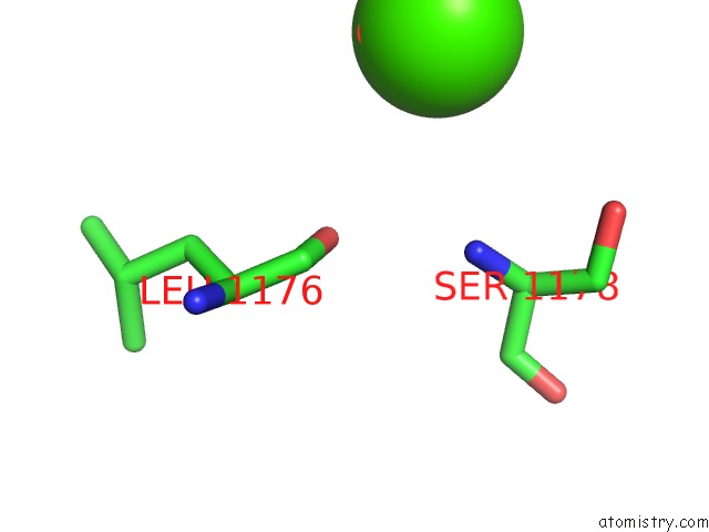
Mono view
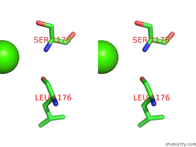
Stereo pair view
Mono view
Stereo pair view
A full contact list of Calcium with other atoms in the Ca binding
site number 1 of Crystal Structure of Voltage-Gated Sodium Channel Navab in High-pH Condition within 5.0Å range:
|
Reference:
K.Irie,
Y.Haga,
T.Shimomura,
Y.Fujiyoshi.
Structural Insight on the Voltage Dependence of Prokaryotic Voltage Gated Sodium Channel Navab. Febs Lett. 2017.
ISSN: ISSN 1873-3468
PubMed: 29274127
DOI: 10.1002/1873-3468.12955
Page generated: Wed Jul 9 12:05:02 2025
ISSN: ISSN 1873-3468
PubMed: 29274127
DOI: 10.1002/1873-3468.12955
Last articles
K in 1ME8K in 1ME7
K in 1MAS
K in 1MC5
K in 1MA0
K in 1M90
K in 1M9N
K in 1M6H
K in 1M6W
K in 1M40