Calcium »
PDB 5z4u-5zxh »
5z6o »
Calcium in PDB 5z6o: Crystal Structure of Penicillium Cyclopium Protease
Enzymatic activity of Crystal Structure of Penicillium Cyclopium Protease
All present enzymatic activity of Crystal Structure of Penicillium Cyclopium Protease:
3.4.21.64;
3.4.21.64;
Protein crystallography data
The structure of Crystal Structure of Penicillium Cyclopium Protease, PDB code: 5z6o
was solved by
T.-P.Ko,
S.Koszelak,
J.Ng,
J.Day,
A.Greenwood,
A.Mcpherson,
with X-Ray Crystallography technique. A brief refinement statistics is given in the table below:
| Resolution Low / High (Å) | 22.72 / 1.70 |
| Space group | P 21 21 21 |
| Cell size a, b, c (Å), α, β, γ (°) | 59.223, 61.642, 70.862, 90.00, 90.00, 90.00 |
| R / Rfree (%) | 14.2 / 18.1 |
Calcium Binding Sites:
The binding sites of Calcium atom in the Crystal Structure of Penicillium Cyclopium Protease
(pdb code 5z6o). This binding sites where shown within
5.0 Angstroms radius around Calcium atom.
In total only one binding site of Calcium was determined in the Crystal Structure of Penicillium Cyclopium Protease, PDB code: 5z6o:
In total only one binding site of Calcium was determined in the Crystal Structure of Penicillium Cyclopium Protease, PDB code: 5z6o:
Calcium binding site 1 out of 1 in 5z6o
Go back to
Calcium binding site 1 out
of 1 in the Crystal Structure of Penicillium Cyclopium Protease
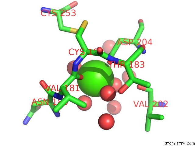
Mono view
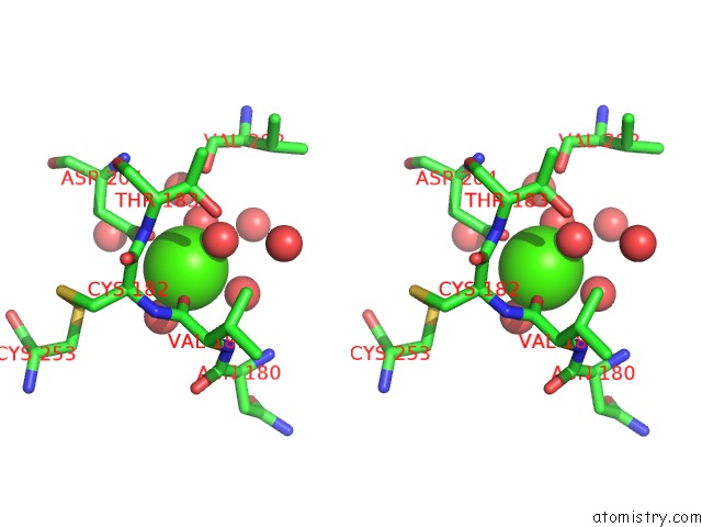
Stereo pair view
Mono view
Stereo pair view
A full contact list of Calcium with other atoms in the Ca binding
site number 1 of Crystal Structure of Penicillium Cyclopium Protease within 5.0Å range:
|
Reference:
S.Koszelak,
J.D.Ng,
J.Day,
T.P.Ko,
A.Greenwood,
A.Mcpherson.
The Crystallographic Structure of the Subtilisin Protease From Penicillium Cyclopium. Biochemistry V. 36 6597 1997.
ISSN: ISSN 0006-2960
PubMed: 9184139
DOI: 10.1021/BI963189T
Page generated: Wed Jul 9 12:11:20 2025
ISSN: ISSN 0006-2960
PubMed: 9184139
DOI: 10.1021/BI963189T
Last articles
Mg in 5IMIMg in 5IMN
Mg in 5ILY
Mg in 5IM3
Mg in 5IK2
Mg in 5ILI
Mg in 5ILG
Mg in 5ILD
Mg in 5IL3
Mg in 5IKZ