Calcium »
PDB 5z4u-5zxh »
5ztf »
Calcium in PDB 5ztf: Structure of CA2+ Atpase
Enzymatic activity of Structure of CA2+ Atpase
All present enzymatic activity of Structure of CA2+ Atpase:
3.6.3.8;
3.6.3.8;
Protein crystallography data
The structure of Structure of CA2+ Atpase, PDB code: 5ztf
was solved by
M.Inoue,
S.Watanabe,
K.Inaba,
with X-Ray Crystallography technique. A brief refinement statistics is given in the table below:
| Resolution Low / High (Å) | 48.22 / 3.45 |
| Space group | P 21 21 2 |
| Cell size a, b, c (Å), α, β, γ (°) | 165.079, 84.053, 118.832, 90.00, 90.00, 90.00 |
| R / Rfree (%) | 24.6 / 30.8 |
Other elements in 5ztf:
The structure of Structure of CA2+ Atpase also contains other interesting chemical elements:
| Magnesium | (Mg) | 1 atom |
Calcium Binding Sites:
The binding sites of Calcium atom in the Structure of CA2+ Atpase
(pdb code 5ztf). This binding sites where shown within
5.0 Angstroms radius around Calcium atom.
In total 2 binding sites of Calcium where determined in the Structure of CA2+ Atpase, PDB code: 5ztf:
Jump to Calcium binding site number: 1; 2;
In total 2 binding sites of Calcium where determined in the Structure of CA2+ Atpase, PDB code: 5ztf:
Jump to Calcium binding site number: 1; 2;
Calcium binding site 1 out of 2 in 5ztf
Go back to
Calcium binding site 1 out
of 2 in the Structure of CA2+ Atpase
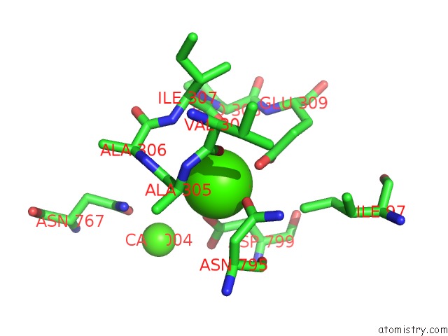
Mono view
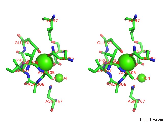
Stereo pair view
Mono view
Stereo pair view
A full contact list of Calcium with other atoms in the Ca binding
site number 1 of Structure of CA2+ Atpase within 5.0Å range:
|
Calcium binding site 2 out of 2 in 5ztf
Go back to
Calcium binding site 2 out
of 2 in the Structure of CA2+ Atpase
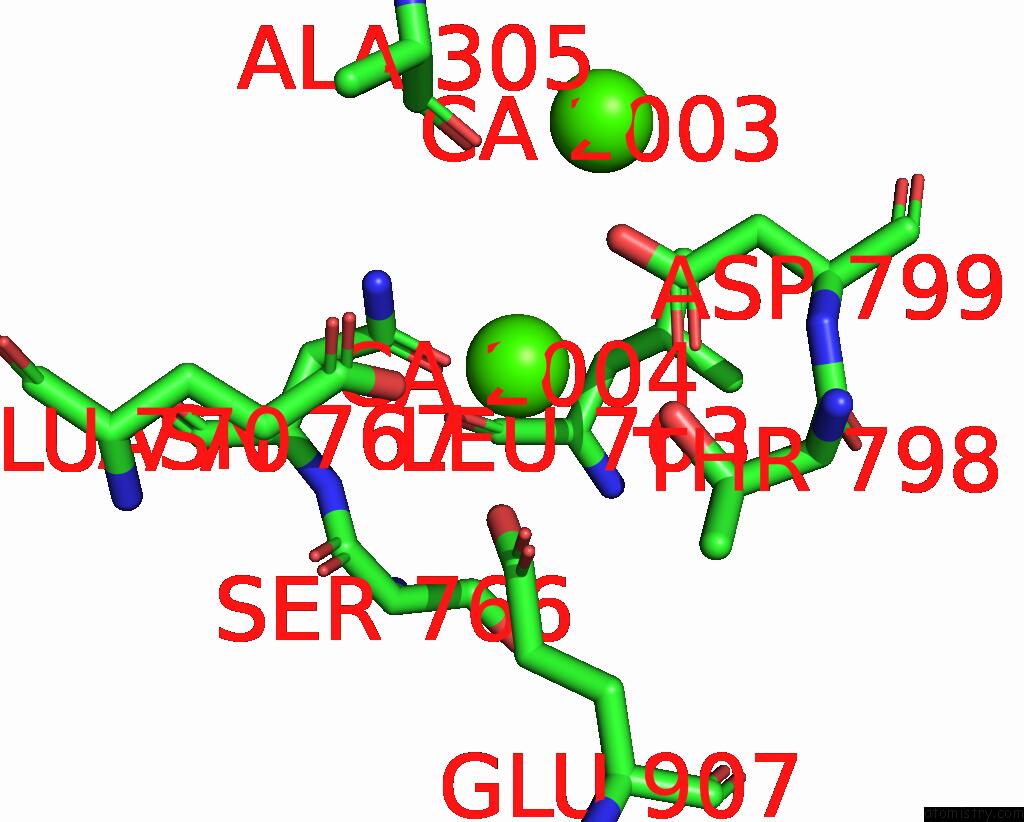
Mono view
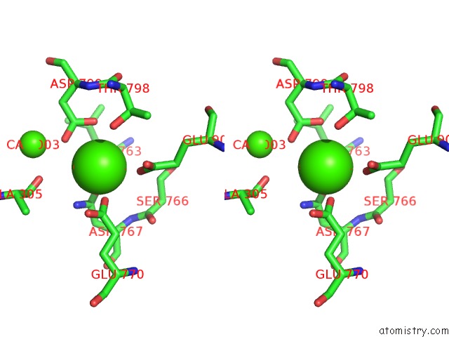
Stereo pair view
Mono view
Stereo pair view
A full contact list of Calcium with other atoms in the Ca binding
site number 2 of Structure of CA2+ Atpase within 5.0Å range:
|
Reference:
M.Inoue,
N.Sakuta,
S.Watanabe,
Y.Zhang,
K.Yoshikaie,
Y.Tanaka,
R.Ushioda,
Y.Kato,
J.Takagi,
T.Tsukazaki,
K.Nagata,
K.Inaba.
Structural Basis of Sarco/Endoplasmic Reticulum CA2+-Atpase 2B Regulation Via Transmembrane Helix Interplay. Cell Rep V. 27 1221 2019.
ISSN: ESSN 2211-1247
PubMed: 31018135
DOI: 10.1016/J.CELREP.2019.03.106
Page generated: Wed Jul 9 12:17:42 2025
ISSN: ESSN 2211-1247
PubMed: 31018135
DOI: 10.1016/J.CELREP.2019.03.106
Last articles
Mg in 1JB0Mg in 1IZL
Mg in 1JBZ
Mg in 1JBW
Mg in 1JBV
Mg in 1JBK
Mg in 1JAX
Mg in 1JAH
Mg in 1J97
Mg in 1J9J