Calcium »
PDB 6b5v-6bia »
6bdt »
Calcium in PDB 6bdt: Crystal Structure of Human Calpain-3 Protease Core Mutant-C129S
Enzymatic activity of Crystal Structure of Human Calpain-3 Protease Core Mutant-C129S
All present enzymatic activity of Crystal Structure of Human Calpain-3 Protease Core Mutant-C129S:
3.4.22.54;
3.4.22.54;
Protein crystallography data
The structure of Crystal Structure of Human Calpain-3 Protease Core Mutant-C129S, PDB code: 6bdt
was solved by
Q.Ye,
R.L.Campbell,
P.L.Davies,
with X-Ray Crystallography technique. A brief refinement statistics is given in the table below:
| Resolution Low / High (Å) | 17.99 / 2.30 |
| Space group | P 21 21 21 |
| Cell size a, b, c (Å), α, β, γ (°) | 59.810, 105.490, 225.260, 90.00, 90.00, 90.00 |
| R / Rfree (%) | 22.2 / 25.6 |
Other elements in 6bdt:
The structure of Crystal Structure of Human Calpain-3 Protease Core Mutant-C129S also contains other interesting chemical elements:
| Chlorine | (Cl) | 4 atoms |
Calcium Binding Sites:
The binding sites of Calcium atom in the Crystal Structure of Human Calpain-3 Protease Core Mutant-C129S
(pdb code 6bdt). This binding sites where shown within
5.0 Angstroms radius around Calcium atom.
In total 8 binding sites of Calcium where determined in the Crystal Structure of Human Calpain-3 Protease Core Mutant-C129S, PDB code: 6bdt:
Jump to Calcium binding site number: 1; 2; 3; 4; 5; 6; 7; 8;
In total 8 binding sites of Calcium where determined in the Crystal Structure of Human Calpain-3 Protease Core Mutant-C129S, PDB code: 6bdt:
Jump to Calcium binding site number: 1; 2; 3; 4; 5; 6; 7; 8;
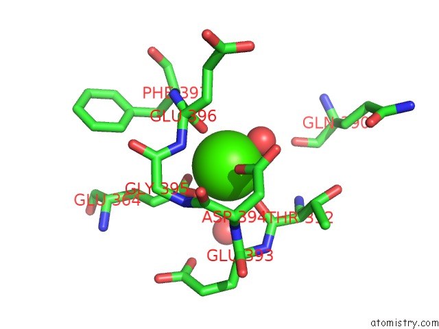
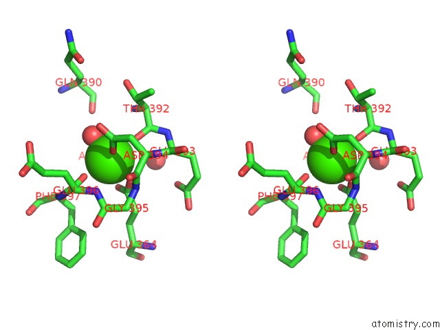
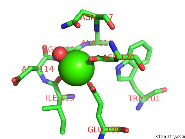
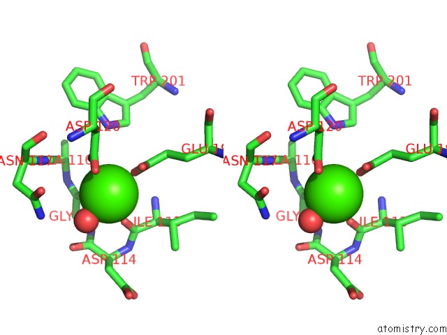
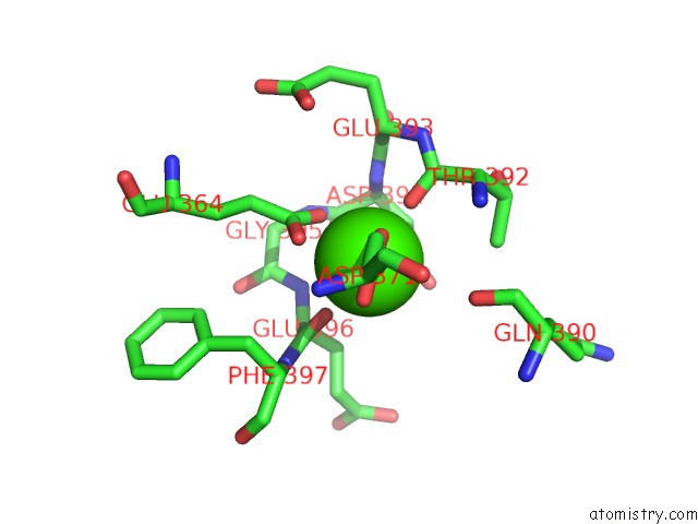
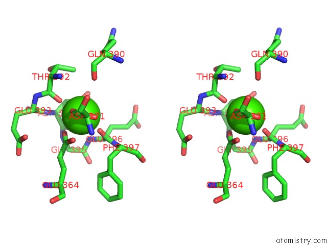
Calcium binding site 1 out of 8 in 6bdt
Go back to
Calcium binding site 1 out
of 8 in the Crystal Structure of Human Calpain-3 Protease Core Mutant-C129S
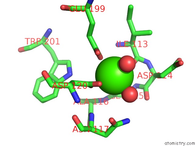
Mono view
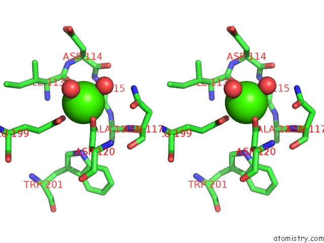
Stereo pair view
Mono view
Stereo pair view
A full contact list of Calcium with other atoms in the Ca binding
site number 1 of Crystal Structure of Human Calpain-3 Protease Core Mutant-C129S within 5.0Å range:
|
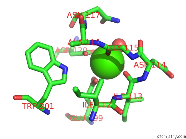
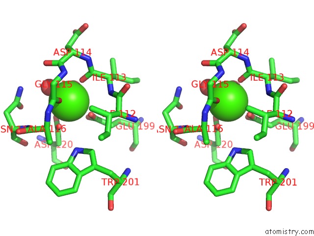
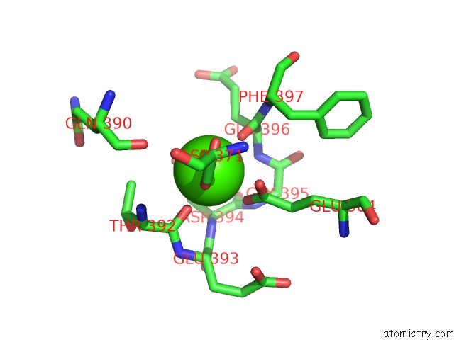
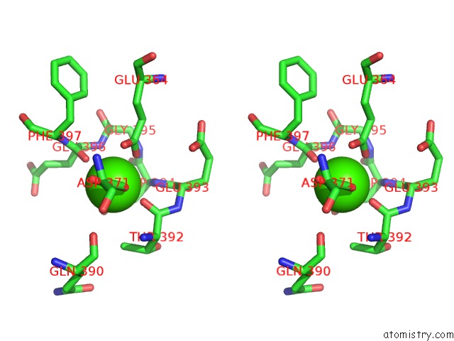
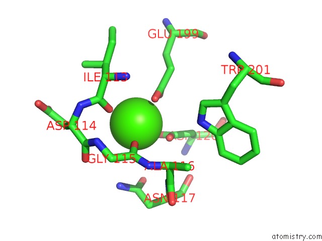
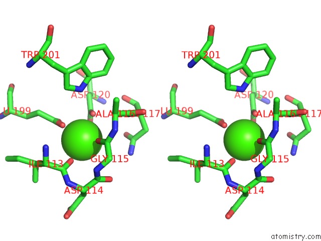
Calcium binding site 2 out of 8 in 6bdt
Go back to
Calcium binding site 2 out
of 8 in the Crystal Structure of Human Calpain-3 Protease Core Mutant-C129S
Mono view
Stereo pair view
Mono view
Stereo pair view
A full contact list of Calcium with other atoms in the Ca binding
site number 2 of Crystal Structure of Human Calpain-3 Protease Core Mutant-C129S within 5.0Å range:
|
Calcium binding site 3 out of 8 in 6bdt
Go back to
Calcium binding site 3 out
of 8 in the Crystal Structure of Human Calpain-3 Protease Core Mutant-C129S
Mono view
Stereo pair view
Mono view
Stereo pair view
A full contact list of Calcium with other atoms in the Ca binding
site number 3 of Crystal Structure of Human Calpain-3 Protease Core Mutant-C129S within 5.0Å range:
|
Calcium binding site 4 out of 8 in 6bdt
Go back to
Calcium binding site 4 out
of 8 in the Crystal Structure of Human Calpain-3 Protease Core Mutant-C129S
Mono view
Stereo pair view
Mono view
Stereo pair view
A full contact list of Calcium with other atoms in the Ca binding
site number 4 of Crystal Structure of Human Calpain-3 Protease Core Mutant-C129S within 5.0Å range:
|
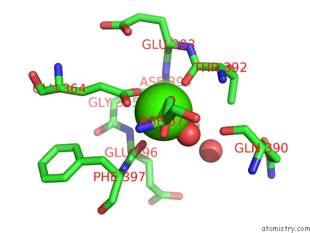
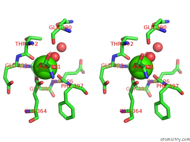
Calcium binding site 5 out of 8 in 6bdt
Go back to
Calcium binding site 5 out
of 8 in the Crystal Structure of Human Calpain-3 Protease Core Mutant-C129S
Mono view
Stereo pair view
Mono view
Stereo pair view
A full contact list of Calcium with other atoms in the Ca binding
site number 5 of Crystal Structure of Human Calpain-3 Protease Core Mutant-C129S within 5.0Å range:
|
Calcium binding site 6 out of 8 in 6bdt
Go back to
Calcium binding site 6 out
of 8 in the Crystal Structure of Human Calpain-3 Protease Core Mutant-C129S
Mono view
Stereo pair view
Mono view
Stereo pair view
A full contact list of Calcium with other atoms in the Ca binding
site number 6 of Crystal Structure of Human Calpain-3 Protease Core Mutant-C129S within 5.0Å range:
|
Calcium binding site 7 out of 8 in 6bdt
Go back to
Calcium binding site 7 out
of 8 in the Crystal Structure of Human Calpain-3 Protease Core Mutant-C129S
Mono view
Stereo pair view
Mono view
Stereo pair view
A full contact list of Calcium with other atoms in the Ca binding
site number 7 of Crystal Structure of Human Calpain-3 Protease Core Mutant-C129S within 5.0Å range:
|
Calcium binding site 8 out of 8 in 6bdt
Go back to
Calcium binding site 8 out
of 8 in the Crystal Structure of Human Calpain-3 Protease Core Mutant-C129S
Mono view
Stereo pair view
Mono view
Stereo pair view
A full contact list of Calcium with other atoms in the Ca binding
site number 8 of Crystal Structure of Human Calpain-3 Protease Core Mutant-C129S within 5.0Å range:
|
Reference:
Q.Ye,
R.L.Campbell,
P.L.Davies.
Structures of Human Calpain-3 Protease Core with and Without Bound Inhibitor Reveal Mechanisms of Calpain Activation. J. Biol. Chem. V. 293 4056 2018.
ISSN: ESSN 1083-351X
PubMed: 29382717
DOI: 10.1074/JBC.RA117.001097
Page generated: Wed Jul 9 12:44:40 2025
ISSN: ESSN 1083-351X
PubMed: 29382717
DOI: 10.1074/JBC.RA117.001097
Last articles
Fe in 7R2OFe in 7R1J
Fe in 7R1I
Fe in 7QWT
Fe in 7R1H
Fe in 7R0F
Fe in 7QZH
Fe in 7QZR
Fe in 7QZG
Fe in 7QZF