Calcium »
PDB 6b5v-6bia »
6bgd »
Calcium in PDB 6bgd: The Crystal Structure of the W145A Variant of Tpmglb-2 (TP0684) with Bound Glucose
Protein crystallography data
The structure of The Crystal Structure of the W145A Variant of Tpmglb-2 (TP0684) with Bound Glucose, PDB code: 6bgd
was solved by
C.A.Brautigam,
M.V.Norgard,
R.K.Deka,
with X-Ray Crystallography technique. A brief refinement statistics is given in the table below:
| Resolution Low / High (Å) | 37.90 / 1.47 |
| Space group | C 2 2 21 |
| Cell size a, b, c (Å), α, β, γ (°) | 75.834, 110.199, 89.304, 90.00, 90.00, 90.00 |
| R / Rfree (%) | 11.4 / 14.4 |
Calcium Binding Sites:
The binding sites of Calcium atom in the The Crystal Structure of the W145A Variant of Tpmglb-2 (TP0684) with Bound Glucose
(pdb code 6bgd). This binding sites where shown within
5.0 Angstroms radius around Calcium atom.
In total only one binding site of Calcium was determined in the The Crystal Structure of the W145A Variant of Tpmglb-2 (TP0684) with Bound Glucose, PDB code: 6bgd:
In total only one binding site of Calcium was determined in the The Crystal Structure of the W145A Variant of Tpmglb-2 (TP0684) with Bound Glucose, PDB code: 6bgd:
Calcium binding site 1 out of 1 in 6bgd
Go back to
Calcium binding site 1 out
of 1 in the The Crystal Structure of the W145A Variant of Tpmglb-2 (TP0684) with Bound Glucose
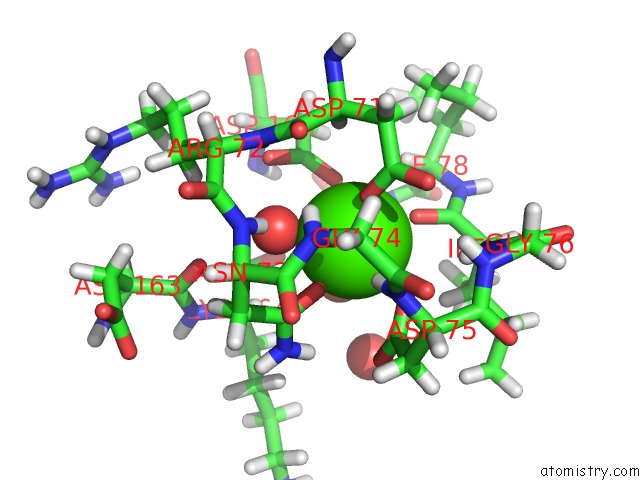
Mono view
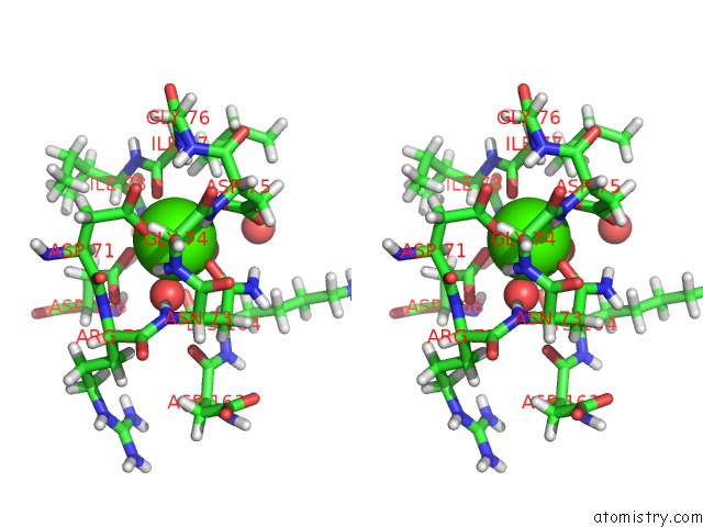
Stereo pair view
Mono view
Stereo pair view
A full contact list of Calcium with other atoms in the Ca binding
site number 1 of The Crystal Structure of the W145A Variant of Tpmglb-2 (TP0684) with Bound Glucose within 5.0Å range:
|
Reference:
C.A.Brautigam,
R.K.Deka,
W.Z.Liu,
M.V.Norgard.
Crystal Structures of Mglb-2 (TP0684), A Topologically Variant D-Glucose-Binding Protein From Treponema Pallidum, Reveal A Ligand-Induced Conformational Change. Protein Sci. V. 27 880 2018.
ISSN: ESSN 1469-896X
PubMed: 29318719
DOI: 10.1002/PRO.3373
Page generated: Wed Jul 9 12:47:17 2025
ISSN: ESSN 1469-896X
PubMed: 29318719
DOI: 10.1002/PRO.3373
Last articles
Mg in 3B9BMg in 3B8E
Mg in 3B7L
Mg in 3B6B
Mg in 3B7W
Mg in 3B6V
Mg in 3B6U
Mg in 3B5I
Mg in 3B4A
Mg in 3B6R