Calcium »
PDB 6bjd-6bxz »
6bst »
Calcium in PDB 6bst: Crystal Structure of Z-Dna with Untypically Coordinated CA2+ Ion
Protein crystallography data
The structure of Crystal Structure of Z-Dna with Untypically Coordinated CA2+ Ion, PDB code: 6bst
was solved by
Z.Luo,
Z.Dauter,
with X-Ray Crystallography technique. A brief refinement statistics is given in the table below:
| Resolution Low / High (Å) | 24.00 / 1.45 |
| Space group | P 21 21 21 |
| Cell size a, b, c (Å), α, β, γ (°) | 17.760, 28.820, 42.360, 90.00, 90.00, 90.00 |
| R / Rfree (%) | 12.1 / 17.2 |
Calcium Binding Sites:
The binding sites of Calcium atom in the Crystal Structure of Z-Dna with Untypically Coordinated CA2+ Ion
(pdb code 6bst). This binding sites where shown within
5.0 Angstroms radius around Calcium atom.
In total only one binding site of Calcium was determined in the Crystal Structure of Z-Dna with Untypically Coordinated CA2+ Ion, PDB code: 6bst:
In total only one binding site of Calcium was determined in the Crystal Structure of Z-Dna with Untypically Coordinated CA2+ Ion, PDB code: 6bst:
Calcium binding site 1 out of 1 in 6bst
Go back to
Calcium binding site 1 out
of 1 in the Crystal Structure of Z-Dna with Untypically Coordinated CA2+ Ion
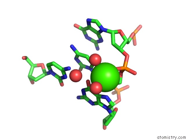
Mono view
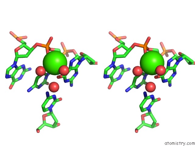
Stereo pair view
Mono view
Stereo pair view
A full contact list of Calcium with other atoms in the Ca binding
site number 1 of Crystal Structure of Z-Dna with Untypically Coordinated CA2+ Ion within 5.0Å range:
|
Reference:
Z.Luo,
Z.Dauter.
The Crystal Structure of Z-Dna with Untypically Coordinated Ca J. Biol. Inorg. Chem. V. 23 253 2018.
ISSN: ESSN 1432-1327
PubMed: 29270817
DOI: 10.1007/S00775-017-1526-4
Page generated: Wed Jul 9 12:53:08 2025
ISSN: ESSN 1432-1327
PubMed: 29270817
DOI: 10.1007/S00775-017-1526-4
Last articles
Mg in 3AQ4Mg in 3ALO
Mg in 3AO2
Mg in 3AM1
Mg in 3AK8
Mg in 3ALN
Mg in 3AKL
Mg in 3AKM
Mg in 3AL5
Mg in 3AKK