Calcium »
PDB 6dmn-6e48 »
6drj »
Calcium in PDB 6drj: Structure of TRPM2 Ion Channel Receptor By Single Particle Electron Cryo-Microscopy, Adpr/CA2+ Bound State
Calcium Binding Sites:
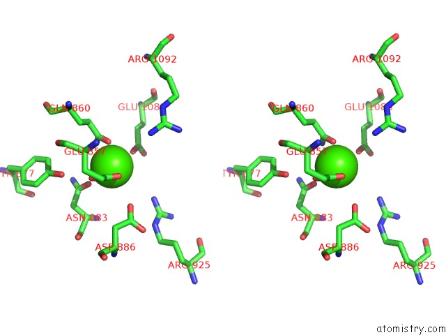
The binding sites of Calcium atom in the Structure of TRPM2 Ion Channel Receptor By Single Particle Electron Cryo-Microscopy, Adpr/CA2+ Bound State
(pdb code 6drj). This binding sites where shown within
5.0 Angstroms radius around Calcium atom.
In total 4 binding sites of Calcium where determined in the Structure of TRPM2 Ion Channel Receptor By Single Particle Electron Cryo-Microscopy, Adpr/CA2+ Bound State, PDB code: 6drj:
Jump to Calcium binding site number: 1; 2; 3; 4;
In total 4 binding sites of Calcium where determined in the Structure of TRPM2 Ion Channel Receptor By Single Particle Electron Cryo-Microscopy, Adpr/CA2+ Bound State, PDB code: 6drj:
Jump to Calcium binding site number: 1; 2; 3; 4;
Calcium binding site 1 out of 4 in 6drj
Go back to
Calcium binding site 1 out
of 4 in the Structure of TRPM2 Ion Channel Receptor By Single Particle Electron Cryo-Microscopy, Adpr/CA2+ Bound State
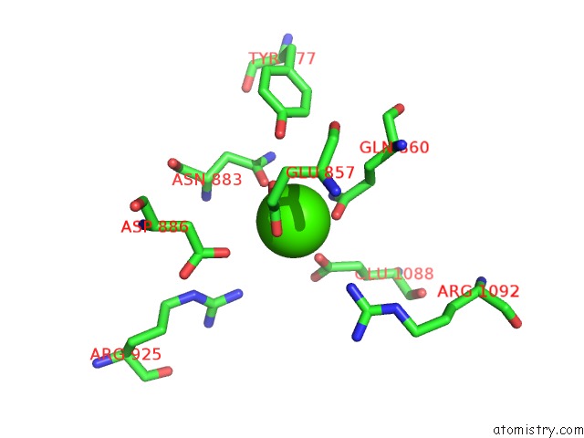
Mono view
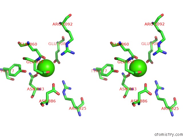
Stereo pair view
Mono view
Stereo pair view
A full contact list of Calcium with other atoms in the Ca binding
site number 1 of Structure of TRPM2 Ion Channel Receptor By Single Particle Electron Cryo-Microscopy, Adpr/CA2+ Bound State within 5.0Å range:
|
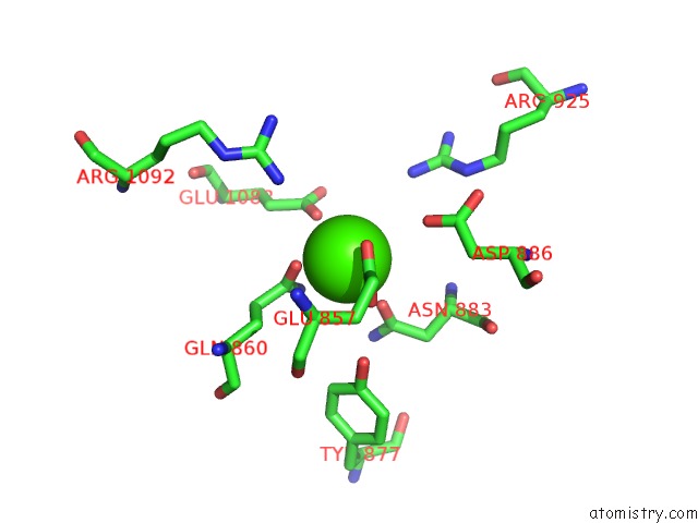
Calcium binding site 2 out of 4 in 6drj
Go back to
Calcium binding site 2 out
of 4 in the Structure of TRPM2 Ion Channel Receptor By Single Particle Electron Cryo-Microscopy, Adpr/CA2+ Bound State

Mono view
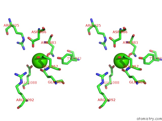
Stereo pair view
Mono view
Stereo pair view
A full contact list of Calcium with other atoms in the Ca binding
site number 2 of Structure of TRPM2 Ion Channel Receptor By Single Particle Electron Cryo-Microscopy, Adpr/CA2+ Bound State within 5.0Å range:
|
Calcium binding site 3 out of 4 in 6drj
Go back to
Calcium binding site 3 out
of 4 in the Structure of TRPM2 Ion Channel Receptor By Single Particle Electron Cryo-Microscopy, Adpr/CA2+ Bound State
Mono view

Stereo pair view
Mono view
Stereo pair view
A full contact list of Calcium with other atoms in the Ca binding
site number 3 of Structure of TRPM2 Ion Channel Receptor By Single Particle Electron Cryo-Microscopy, Adpr/CA2+ Bound State within 5.0Å range:
|
Calcium binding site 4 out of 4 in 6drj
Go back to
Calcium binding site 4 out
of 4 in the Structure of TRPM2 Ion Channel Receptor By Single Particle Electron Cryo-Microscopy, Adpr/CA2+ Bound State

Mono view
Stereo pair view
Mono view
Stereo pair view
A full contact list of Calcium with other atoms in the Ca binding
site number 4 of Structure of TRPM2 Ion Channel Receptor By Single Particle Electron Cryo-Microscopy, Adpr/CA2+ Bound State within 5.0Å range:
|
Reference:
Y.Huang,
P.A.Winkler,
W.Sun,
W.Lu,
J.Du.
Architecture of the TRPM2 Channel and Its Activation Mechanism By Adp-Ribose and Calcium. Nature V. 562 145 2018.
ISSN: ISSN 0028-0836
PubMed: 30250252
DOI: 10.1038/S41586-018-0558-4
Page generated: Wed Jul 9 13:28:11 2025
ISSN: ISSN 0028-0836
PubMed: 30250252
DOI: 10.1038/S41586-018-0558-4
Last articles
Mg in 5ZKJMg in 5ZKI
Mg in 5ZK6
Mg in 5ZE9
Mg in 5ZFX
Mg in 5ZCT
Mg in 5ZE6
Mg in 5ZE4
Mg in 5ZDN
Mg in 5ZE0