Calcium »
PDB 6e54-6ela »
6e54 »
Calcium in PDB 6e54: Crystal Structure of Lpxc From Pseudomonas Aeruginosa in Complex with Ligand PT802
Enzymatic activity of Crystal Structure of Lpxc From Pseudomonas Aeruginosa in Complex with Ligand PT802
All present enzymatic activity of Crystal Structure of Lpxc From Pseudomonas Aeruginosa in Complex with Ligand PT802:
3.5.1.108;
3.5.1.108;
Protein crystallography data
The structure of Crystal Structure of Lpxc From Pseudomonas Aeruginosa in Complex with Ligand PT802, PDB code: 6e54
was solved by
Seattle Structural Genomics Center For Infectious Disease,
Seattlestructural Genomics Center For Infectious Disease (Ssgcid),
with X-Ray Crystallography technique. A brief refinement statistics is given in the table below:
| Resolution Low / High (Å) | 39.58 / 1.65 |
| Space group | P 1 |
| Cell size a, b, c (Å), α, β, γ (°) | 35.640, 47.740, 48.350, 111.68, 107.87, 99.27 |
| R / Rfree (%) | 14.9 / 18.5 |
Other elements in 6e54:
The structure of Crystal Structure of Lpxc From Pseudomonas Aeruginosa in Complex with Ligand PT802 also contains other interesting chemical elements:
| Fluorine | (F) | 2 atoms |
| Zinc | (Zn) | 1 atom |
| Chlorine | (Cl) | 2 atoms |
Calcium Binding Sites:
The binding sites of Calcium atom in the Crystal Structure of Lpxc From Pseudomonas Aeruginosa in Complex with Ligand PT802
(pdb code 6e54). This binding sites where shown within
5.0 Angstroms radius around Calcium atom.
In total 2 binding sites of Calcium where determined in the Crystal Structure of Lpxc From Pseudomonas Aeruginosa in Complex with Ligand PT802, PDB code: 6e54:
Jump to Calcium binding site number: 1; 2;
In total 2 binding sites of Calcium where determined in the Crystal Structure of Lpxc From Pseudomonas Aeruginosa in Complex with Ligand PT802, PDB code: 6e54:
Jump to Calcium binding site number: 1; 2;
Calcium binding site 1 out of 2 in 6e54
Go back to
Calcium binding site 1 out
of 2 in the Crystal Structure of Lpxc From Pseudomonas Aeruginosa in Complex with Ligand PT802
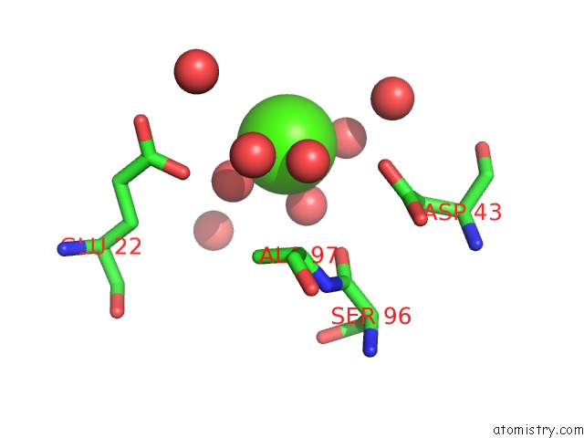
Mono view
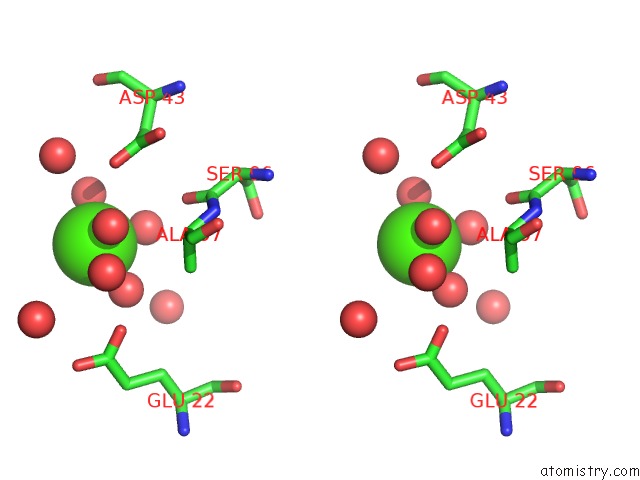
Stereo pair view
Mono view
Stereo pair view
A full contact list of Calcium with other atoms in the Ca binding
site number 1 of Crystal Structure of Lpxc From Pseudomonas Aeruginosa in Complex with Ligand PT802 within 5.0Å range:
|
Calcium binding site 2 out of 2 in 6e54
Go back to
Calcium binding site 2 out
of 2 in the Crystal Structure of Lpxc From Pseudomonas Aeruginosa in Complex with Ligand PT802
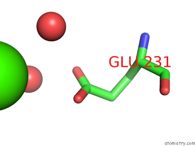
Mono view
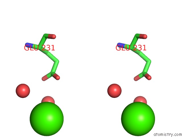
Stereo pair view
Mono view
Stereo pair view
A full contact list of Calcium with other atoms in the Ca binding
site number 2 of Crystal Structure of Lpxc From Pseudomonas Aeruginosa in Complex with Ligand PT802 within 5.0Å range:
|
Reference:
S.L.Delker,
S.J.Mayclin,
J.N.Phan,
J.Abendroth,
D.Lorimer,
T.E.Edwards.
Crystal Structure of Lpxc From Pseudomonas Aeruginosa in Complex with Ligand PT802 To Be Published.
Page generated: Wed Jul 9 13:34:39 2025
Last articles
Mg in 7DSJMg in 7DSI
Mg in 7DRP
Mg in 7DSH
Mg in 7DSA
Mg in 7DRX
Mg in 7DRM
Mg in 7DPT
Mg in 7DQE
Mg in 7DQD