Calcium »
PDB 6elt-6f2y »
6epa »
Calcium in PDB 6epa: Structure of Dncs-1 Bound to the Ncs-1/RIC8A Protein/Protein Interaction Regulator Igs-1.76
Protein crystallography data
The structure of Structure of Dncs-1 Bound to the Ncs-1/RIC8A Protein/Protein Interaction Regulator Igs-1.76, PDB code: 6epa
was solved by
M.J.Sanchez-Barrena,
M.Daniel,
L.Infantes,
with X-Ray Crystallography technique. A brief refinement statistics is given in the table below:
| Resolution Low / High (Å) | 41.11 / 1.82 |
| Space group | P 21 21 21 |
| Cell size a, b, c (Å), α, β, γ (°) | 54.843, 55.456, 61.259, 90.00, 90.00, 90.00 |
| R / Rfree (%) | 18.9 / 23.2 |
Other elements in 6epa:
The structure of Structure of Dncs-1 Bound to the Ncs-1/RIC8A Protein/Protein Interaction Regulator Igs-1.76 also contains other interesting chemical elements:
| Sodium | (Na) | 1 atom |
Calcium Binding Sites:
The binding sites of Calcium atom in the Structure of Dncs-1 Bound to the Ncs-1/RIC8A Protein/Protein Interaction Regulator Igs-1.76
(pdb code 6epa). This binding sites where shown within
5.0 Angstroms radius around Calcium atom.
In total 3 binding sites of Calcium where determined in the Structure of Dncs-1 Bound to the Ncs-1/RIC8A Protein/Protein Interaction Regulator Igs-1.76, PDB code: 6epa:
Jump to Calcium binding site number: 1; 2; 3;
In total 3 binding sites of Calcium where determined in the Structure of Dncs-1 Bound to the Ncs-1/RIC8A Protein/Protein Interaction Regulator Igs-1.76, PDB code: 6epa:
Jump to Calcium binding site number: 1; 2; 3;
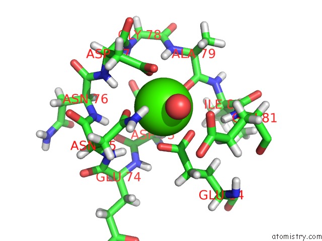
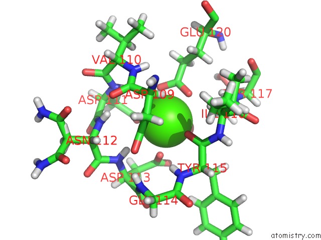
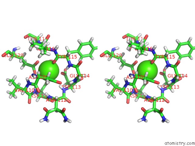
Calcium binding site 1 out of 3 in 6epa
Go back to
Calcium binding site 1 out
of 3 in the Structure of Dncs-1 Bound to the Ncs-1/RIC8A Protein/Protein Interaction Regulator Igs-1.76
Mono view
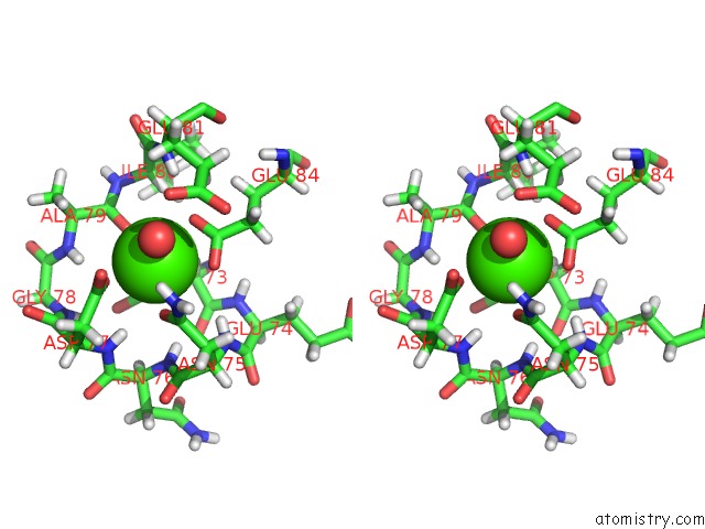
Stereo pair view
Mono view
Stereo pair view
A full contact list of Calcium with other atoms in the Ca binding
site number 1 of Structure of Dncs-1 Bound to the Ncs-1/RIC8A Protein/Protein Interaction Regulator Igs-1.76 within 5.0Å range:
|
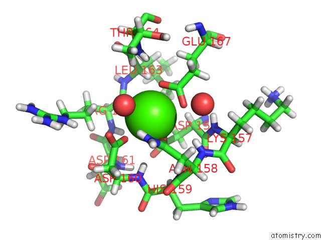
Calcium binding site 2 out of 3 in 6epa
Go back to
Calcium binding site 2 out
of 3 in the Structure of Dncs-1 Bound to the Ncs-1/RIC8A Protein/Protein Interaction Regulator Igs-1.76
Mono view
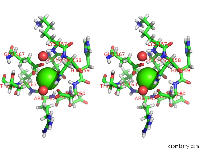
Stereo pair view
Mono view
Stereo pair view
A full contact list of Calcium with other atoms in the Ca binding
site number 2 of Structure of Dncs-1 Bound to the Ncs-1/RIC8A Protein/Protein Interaction Regulator Igs-1.76 within 5.0Å range:
|
Calcium binding site 3 out of 3 in 6epa
Go back to
Calcium binding site 3 out
of 3 in the Structure of Dncs-1 Bound to the Ncs-1/RIC8A Protein/Protein Interaction Regulator Igs-1.76
Mono view
Stereo pair view
Mono view
Stereo pair view
A full contact list of Calcium with other atoms in the Ca binding
site number 3 of Structure of Dncs-1 Bound to the Ncs-1/RIC8A Protein/Protein Interaction Regulator Igs-1.76 within 5.0Å range:
|
Reference:
C.Roca,
L.Martinez-Gonzalez,
M.Daniel-Mozo,
J.Sastre,
L.Infantes,
A.Mansilla,
A.Chaves-Sanjuan,
J.M.Gonzalez-Rubio,
C.Gil,
F.J.Canada,
A.Martinez,
M.J.Sanchez-Barrena,
N.E.Campillo.
Deciphering the Inhibition of the Neuronal Calcium Sensor 1 and the Guanine Exchange Factor RIC8A with A Small Phenothiazine Molecule For the Rational Generation of Therapeutic Synapse Function Regulators. J. Med. Chem. V. 61 5910 2018.
ISSN: ISSN 1520-4804
PubMed: 29966094
DOI: 10.1021/ACS.JMEDCHEM.8B00088
Page generated: Wed Jul 9 13:47:12 2025
ISSN: ISSN 1520-4804
PubMed: 29966094
DOI: 10.1021/ACS.JMEDCHEM.8B00088
Last articles
Mg in 3CRLMg in 3CRR
Mg in 3CRQ
Mg in 3CRC
Mg in 3CR3
Mg in 3CQ3
Mg in 3CQD
Mg in 3CR1
Mg in 3CME
Mg in 3CMA