Calcium »
PDB 6elt-6f2y »
6ew9 »
Calcium in PDB 6ew9: Crystal Structure of Degs Stress Sensor Protease in Complex with Activating Dnrlglvyqf Peptide
Enzymatic activity of Crystal Structure of Degs Stress Sensor Protease in Complex with Activating Dnrlglvyqf Peptide
All present enzymatic activity of Crystal Structure of Degs Stress Sensor Protease in Complex with Activating Dnrlglvyqf Peptide:
3.4.21.107;
3.4.21.107;
Protein crystallography data
The structure of Crystal Structure of Degs Stress Sensor Protease in Complex with Activating Dnrlglvyqf Peptide, PDB code: 6ew9
was solved by
I.R.Vetter,
A.T.Porfetye,
P.Stege,
with X-Ray Crystallography technique. A brief refinement statistics is given in the table below:
| Resolution Low / High (Å) | 43.38 / 2.20 |
| Space group | P 1 2 1 |
| Cell size a, b, c (Å), α, β, γ (°) | 71.600, 53.650, 132.500, 90.00, 101.80, 90.00 |
| R / Rfree (%) | 22.6 / 27.3 |
Calcium Binding Sites:
The binding sites of Calcium atom in the Crystal Structure of Degs Stress Sensor Protease in Complex with Activating Dnrlglvyqf Peptide
(pdb code 6ew9). This binding sites where shown within
5.0 Angstroms radius around Calcium atom.
In total 8 binding sites of Calcium where determined in the Crystal Structure of Degs Stress Sensor Protease in Complex with Activating Dnrlglvyqf Peptide, PDB code: 6ew9:
Jump to Calcium binding site number: 1; 2; 3; 4; 5; 6; 7; 8;
In total 8 binding sites of Calcium where determined in the Crystal Structure of Degs Stress Sensor Protease in Complex with Activating Dnrlglvyqf Peptide, PDB code: 6ew9:
Jump to Calcium binding site number: 1; 2; 3; 4; 5; 6; 7; 8;
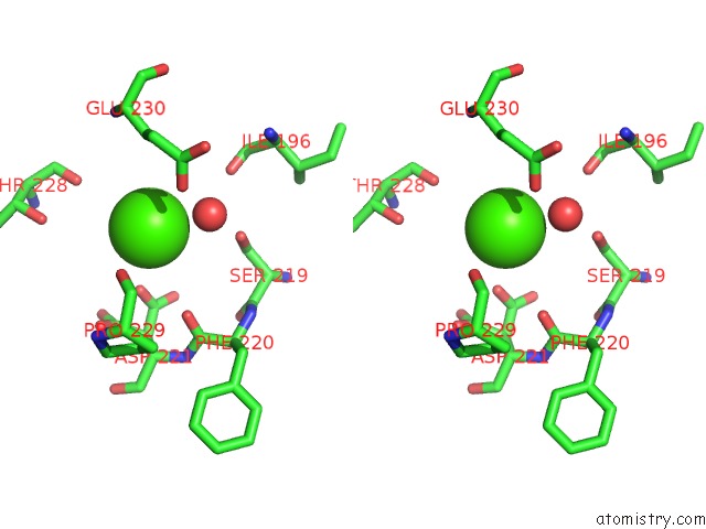
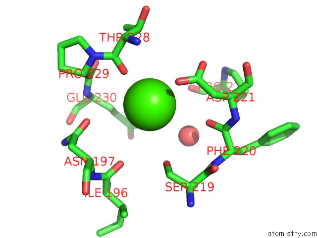
Calcium binding site 1 out of 8 in 6ew9
Go back to
Calcium binding site 1 out
of 8 in the Crystal Structure of Degs Stress Sensor Protease in Complex with Activating Dnrlglvyqf Peptide

Mono view
Stereo pair view
Mono view
Stereo pair view
A full contact list of Calcium with other atoms in the Ca binding
site number 1 of Crystal Structure of Degs Stress Sensor Protease in Complex with Activating Dnrlglvyqf Peptide within 5.0Å range:
|
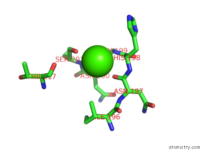
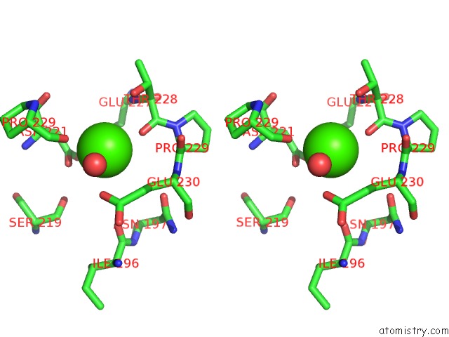
Calcium binding site 2 out of 8 in 6ew9
Go back to
Calcium binding site 2 out
of 8 in the Crystal Structure of Degs Stress Sensor Protease in Complex with Activating Dnrlglvyqf Peptide
Mono view
Stereo pair view
Mono view
Stereo pair view
A full contact list of Calcium with other atoms in the Ca binding
site number 2 of Crystal Structure of Degs Stress Sensor Protease in Complex with Activating Dnrlglvyqf Peptide within 5.0Å range:
|
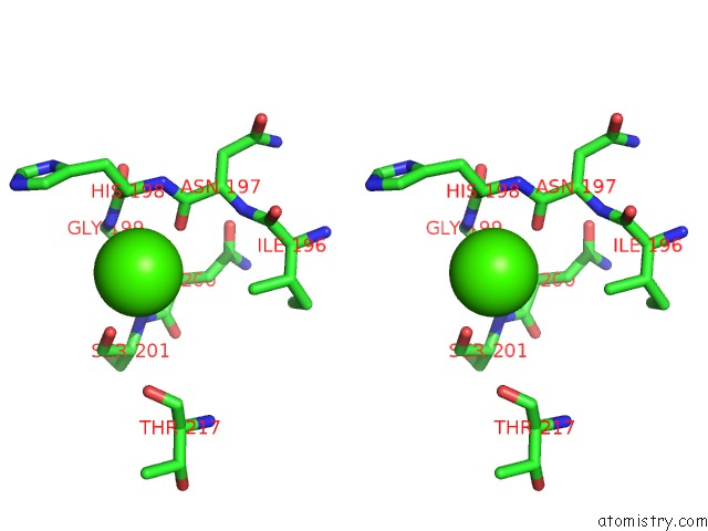
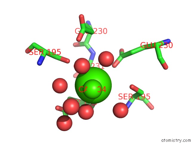
Calcium binding site 3 out of 8 in 6ew9
Go back to
Calcium binding site 3 out
of 8 in the Crystal Structure of Degs Stress Sensor Protease in Complex with Activating Dnrlglvyqf Peptide
Mono view

Stereo pair view
Mono view
Stereo pair view
A full contact list of Calcium with other atoms in the Ca binding
site number 3 of Crystal Structure of Degs Stress Sensor Protease in Complex with Activating Dnrlglvyqf Peptide within 5.0Å range:
|
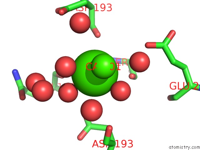
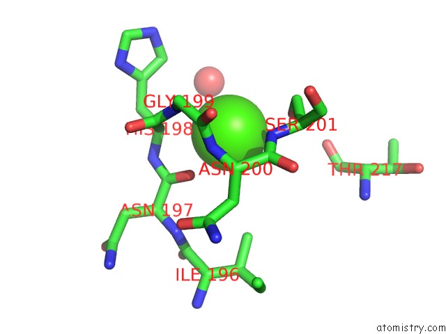
Calcium binding site 4 out of 8 in 6ew9
Go back to
Calcium binding site 4 out
of 8 in the Crystal Structure of Degs Stress Sensor Protease in Complex with Activating Dnrlglvyqf Peptide
Mono view

Stereo pair view
Mono view
Stereo pair view
A full contact list of Calcium with other atoms in the Ca binding
site number 4 of Crystal Structure of Degs Stress Sensor Protease in Complex with Activating Dnrlglvyqf Peptide within 5.0Å range:
|
Calcium binding site 5 out of 8 in 6ew9
Go back to
Calcium binding site 5 out
of 8 in the Crystal Structure of Degs Stress Sensor Protease in Complex with Activating Dnrlglvyqf Peptide

Mono view

Stereo pair view
Mono view
Stereo pair view
A full contact list of Calcium with other atoms in the Ca binding
site number 5 of Crystal Structure of Degs Stress Sensor Protease in Complex with Activating Dnrlglvyqf Peptide within 5.0Å range:
|
Calcium binding site 6 out of 8 in 6ew9
Go back to
Calcium binding site 6 out
of 8 in the Crystal Structure of Degs Stress Sensor Protease in Complex with Activating Dnrlglvyqf Peptide

Mono view
Stereo pair view
Mono view
Stereo pair view
A full contact list of Calcium with other atoms in the Ca binding
site number 6 of Crystal Structure of Degs Stress Sensor Protease in Complex with Activating Dnrlglvyqf Peptide within 5.0Å range:
|
Calcium binding site 7 out of 8 in 6ew9
Go back to
Calcium binding site 7 out
of 8 in the Crystal Structure of Degs Stress Sensor Protease in Complex with Activating Dnrlglvyqf Peptide
Mono view

Stereo pair view
Mono view
Stereo pair view
A full contact list of Calcium with other atoms in the Ca binding
site number 7 of Crystal Structure of Degs Stress Sensor Protease in Complex with Activating Dnrlglvyqf Peptide within 5.0Å range:
|
Calcium binding site 8 out of 8 in 6ew9
Go back to
Calcium binding site 8 out
of 8 in the Crystal Structure of Degs Stress Sensor Protease in Complex with Activating Dnrlglvyqf Peptide
Mono view

Stereo pair view
Mono view
Stereo pair view
A full contact list of Calcium with other atoms in the Ca binding
site number 8 of Crystal Structure of Degs Stress Sensor Protease in Complex with Activating Dnrlglvyqf Peptide within 5.0Å range:
|
Reference:
J.Bongard,
M.Lorenz,
I.R.Vetter,
P.Stege,
A.T.Porfetye,
A.L.Schmitz,
F.Kaschani,
A.Wolf,
U.Koch,
P.Nussbaumer,
B.Klebl,
M.Kaiser,
M.Ehrmann.
Identification of Noncatalytic Lysine Residues From Allosteric Circuits Via Covalent Probes. Acs Chem. Biol. V. 13 1307 2018.
ISSN: ESSN 1554-8937
PubMed: 29658704
DOI: 10.1021/ACSCHEMBIO.8B00101
Page generated: Wed Jul 9 13:54:47 2025
ISSN: ESSN 1554-8937
PubMed: 29658704
DOI: 10.1021/ACSCHEMBIO.8B00101
Last articles
Mg in 6FD4Mg in 6FD5
Mg in 6FD6
Mg in 6FD3
Mg in 6FCT
Mg in 6FCW
Mg in 6FCY
Mg in 6FAI
Mg in 6FCI
Mg in 6FCP