Calcium »
PDB 6fi8-6fzv »
6fwk »
Calcium in PDB 6fwk: The Crystal Structure of POL2CORE-M644G in Complex with Dna and An Incoming Nucleotide
Enzymatic activity of The Crystal Structure of POL2CORE-M644G in Complex with Dna and An Incoming Nucleotide
All present enzymatic activity of The Crystal Structure of POL2CORE-M644G in Complex with Dna and An Incoming Nucleotide:
2.7.7.7;
2.7.7.7;
Protein crystallography data
The structure of The Crystal Structure of POL2CORE-M644G in Complex with Dna and An Incoming Nucleotide, PDB code: 6fwk
was solved by
V.Parkash,
E.Johansson,
with X-Ray Crystallography technique. A brief refinement statistics is given in the table below:
| Resolution Low / High (Å) | 19.99 / 2.50 |
| Space group | P 1 2 1 |
| Cell size a, b, c (Å), α, β, γ (°) | 153.961, 70.342, 158.965, 90.00, 112.82, 90.00 |
| R / Rfree (%) | 22.3 / 26.3 |
Other elements in 6fwk:
The structure of The Crystal Structure of POL2CORE-M644G in Complex with Dna and An Incoming Nucleotide also contains other interesting chemical elements:
| Iron | (Fe) | 2 atoms |
Calcium Binding Sites:
The binding sites of Calcium atom in the The Crystal Structure of POL2CORE-M644G in Complex with Dna and An Incoming Nucleotide
(pdb code 6fwk). This binding sites where shown within
5.0 Angstroms radius around Calcium atom.
In total 5 binding sites of Calcium where determined in the The Crystal Structure of POL2CORE-M644G in Complex with Dna and An Incoming Nucleotide, PDB code: 6fwk:
Jump to Calcium binding site number: 1; 2; 3; 4; 5;
In total 5 binding sites of Calcium where determined in the The Crystal Structure of POL2CORE-M644G in Complex with Dna and An Incoming Nucleotide, PDB code: 6fwk:
Jump to Calcium binding site number: 1; 2; 3; 4; 5;
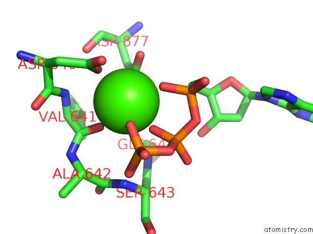
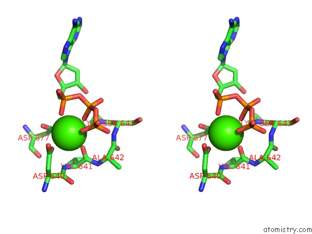
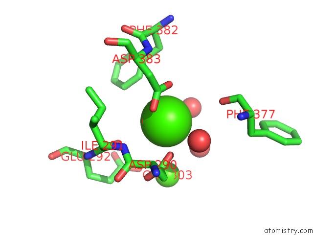
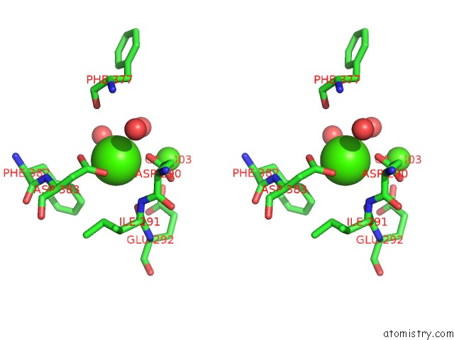
Calcium binding site 1 out of 5 in 6fwk
Go back to
Calcium binding site 1 out
of 5 in the The Crystal Structure of POL2CORE-M644G in Complex with Dna and An Incoming Nucleotide
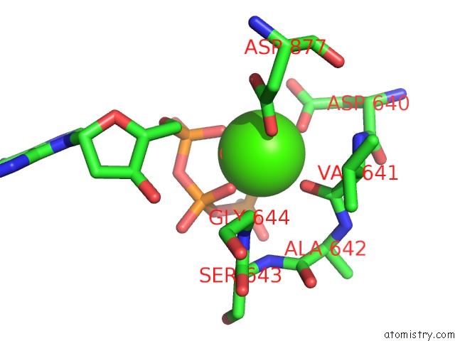
Mono view
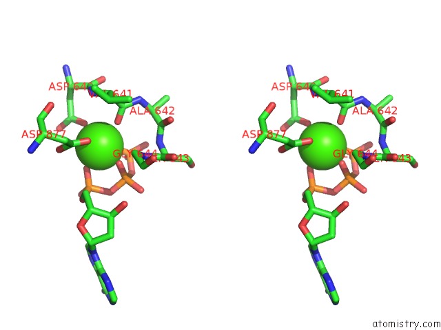
Stereo pair view
Mono view
Stereo pair view
A full contact list of Calcium with other atoms in the Ca binding
site number 1 of The Crystal Structure of POL2CORE-M644G in Complex with Dna and An Incoming Nucleotide within 5.0Å range:
|
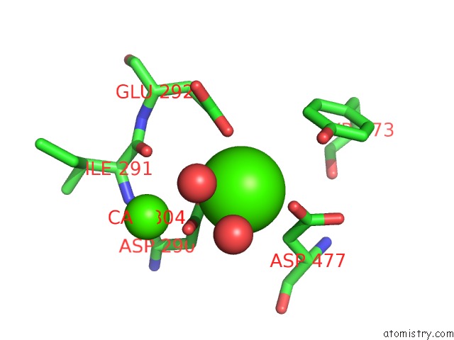
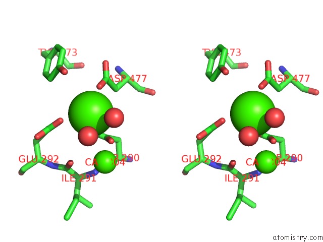
Calcium binding site 2 out of 5 in 6fwk
Go back to
Calcium binding site 2 out
of 5 in the The Crystal Structure of POL2CORE-M644G in Complex with Dna and An Incoming Nucleotide
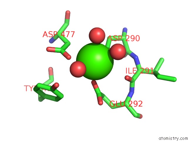
Mono view
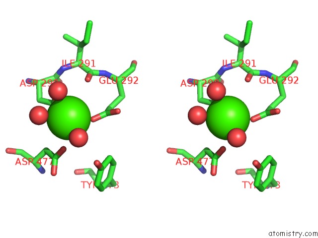
Stereo pair view
Mono view
Stereo pair view
A full contact list of Calcium with other atoms in the Ca binding
site number 2 of The Crystal Structure of POL2CORE-M644G in Complex with Dna and An Incoming Nucleotide within 5.0Å range:
|
Calcium binding site 3 out of 5 in 6fwk
Go back to
Calcium binding site 3 out
of 5 in the The Crystal Structure of POL2CORE-M644G in Complex with Dna and An Incoming Nucleotide
Mono view
Stereo pair view
Mono view
Stereo pair view
A full contact list of Calcium with other atoms in the Ca binding
site number 3 of The Crystal Structure of POL2CORE-M644G in Complex with Dna and An Incoming Nucleotide within 5.0Å range:
|
Calcium binding site 4 out of 5 in 6fwk
Go back to
Calcium binding site 4 out
of 5 in the The Crystal Structure of POL2CORE-M644G in Complex with Dna and An Incoming Nucleotide
Mono view
Stereo pair view
Mono view
Stereo pair view
A full contact list of Calcium with other atoms in the Ca binding
site number 4 of The Crystal Structure of POL2CORE-M644G in Complex with Dna and An Incoming Nucleotide within 5.0Å range:
|
Calcium binding site 5 out of 5 in 6fwk
Go back to
Calcium binding site 5 out
of 5 in the The Crystal Structure of POL2CORE-M644G in Complex with Dna and An Incoming Nucleotide
Mono view
Stereo pair view
Mono view
Stereo pair view
A full contact list of Calcium with other atoms in the Ca binding
site number 5 of The Crystal Structure of POL2CORE-M644G in Complex with Dna and An Incoming Nucleotide within 5.0Å range:
|
Reference:
V.Parkash,
Y.Kulkarni,
J.Ter Beek,
P.V.Shcherbakova,
S.C.L.Kamerlin,
E.Johansson.
Structural Consequence of the Most Frequently Recurring Cancer-Associated Substitution in Dna Polymerase Epsilon. Nat Commun V. 10 373 2019.
ISSN: ESSN 2041-1723
PubMed: 30670696
DOI: 10.1038/S41467-018-08114-9
Page generated: Wed Jul 9 14:18:30 2025
ISSN: ESSN 2041-1723
PubMed: 30670696
DOI: 10.1038/S41467-018-08114-9
Last articles
Mg in 3X0EMg in 3X1L
Mg in 3X1D
Mg in 3WZY
Mg in 3WZX
Mg in 3WYM
Mg in 3WZW
Mg in 3WZV
Mg in 3WYL
Mg in 3WVL