Calcium »
PDB 6fzw-6guy »
6gqg »
Calcium in PDB 6gqg: Structure of GFPMUT2 Crystallized at pH 8.5
Protein crystallography data
The structure of Structure of GFPMUT2 Crystallized at pH 8.5, PDB code: 6gqg
was solved by
E.Pasqualetto,
G.Lolli,
R.Battistutta,
with X-Ray Crystallography technique. A brief refinement statistics is given in the table below:
| Resolution Low / High (Å) | 46.50 / 1.79 |
| Space group | P 21 21 21 |
| Cell size a, b, c (Å), α, β, γ (°) | 51.166, 62.375, 69.767, 90.00, 90.00, 90.00 |
| R / Rfree (%) | 18.3 / 22.3 |
Calcium Binding Sites:
The binding sites of Calcium atom in the Structure of GFPMUT2 Crystallized at pH 8.5
(pdb code 6gqg). This binding sites where shown within
5.0 Angstroms radius around Calcium atom.
In total 2 binding sites of Calcium where determined in the Structure of GFPMUT2 Crystallized at pH 8.5, PDB code: 6gqg:
Jump to Calcium binding site number: 1; 2;
In total 2 binding sites of Calcium where determined in the Structure of GFPMUT2 Crystallized at pH 8.5, PDB code: 6gqg:
Jump to Calcium binding site number: 1; 2;
Calcium binding site 1 out of 2 in 6gqg
Go back to
Calcium binding site 1 out
of 2 in the Structure of GFPMUT2 Crystallized at pH 8.5
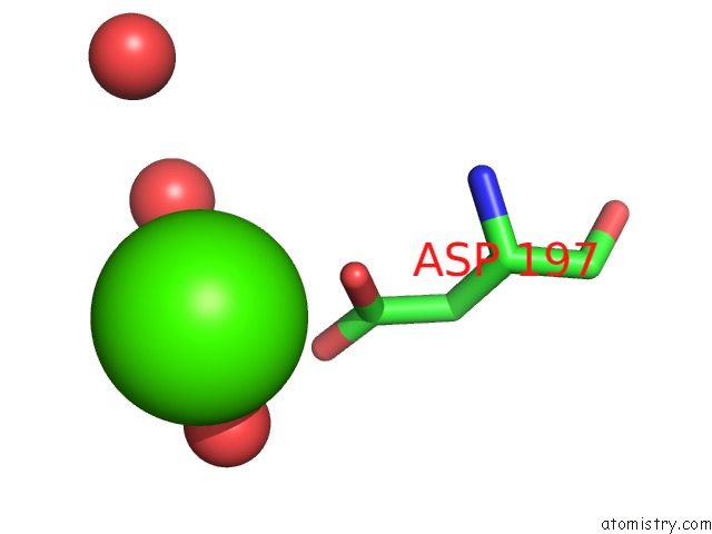
Mono view
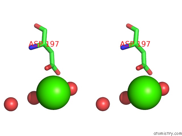
Stereo pair view
Mono view
Stereo pair view
A full contact list of Calcium with other atoms in the Ca binding
site number 1 of Structure of GFPMUT2 Crystallized at pH 8.5 within 5.0Å range:
|
Calcium binding site 2 out of 2 in 6gqg
Go back to
Calcium binding site 2 out
of 2 in the Structure of GFPMUT2 Crystallized at pH 8.5
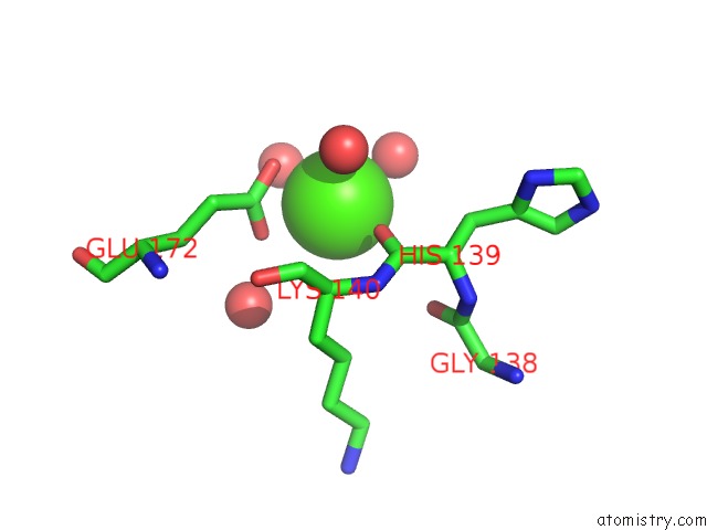
Mono view
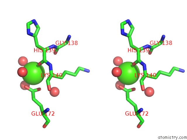
Stereo pair view
Mono view
Stereo pair view
A full contact list of Calcium with other atoms in the Ca binding
site number 2 of Structure of GFPMUT2 Crystallized at pH 8.5 within 5.0Å range:
|
Reference:
G.Lolli,
S.Raboni,
E.Pasqualetto,
R.Benoni,
B.Campanini,
L.Ronda,
A.Mozzarelli,
S.Bettati,
R.Battistutta.
Insight Into GFPMUT2 pH Dependence By Single Crystal Microspectrophotometry and X-Ray Crystallography. J.Phys.Chem.B V. 122 11326 2018.
ISSN: ISSN 1089-5647
PubMed: 30179482
DOI: 10.1021/ACS.JPCB.8B07260
Page generated: Wed Jul 9 14:26:12 2025
ISSN: ISSN 1089-5647
PubMed: 30179482
DOI: 10.1021/ACS.JPCB.8B07260
Last articles
Mg in 5SKEMg in 5SKF
Mg in 5SKD
Mg in 5SKB
Mg in 5SKC
Mg in 5SKA
Mg in 5SK9
Mg in 5SK7
Mg in 5SK6
Mg in 5SK5