Calcium »
PDB 6guz-6hhz »
6har »
Calcium in PDB 6har: Crystal Structure of Mesotrypsin in Complex with Appi-M17C/I18F/F34C
Protein crystallography data
The structure of Crystal Structure of Mesotrypsin in Complex with Appi-M17C/I18F/F34C, PDB code: 6har
was solved by
A.Shahar,
I.Cohen,
E.Radisky,
N.Papo,
with X-Ray Crystallography technique. A brief refinement statistics is given in the table below:
| Resolution Low / High (Å) | 46.47 / 1.50 |
| Space group | P 1 21 1 |
| Cell size a, b, c (Å), α, β, γ (°) | 34.091, 82.781, 46.559, 90.00, 93.63, 90.00 |
| R / Rfree (%) | 16.9 / 19.3 |
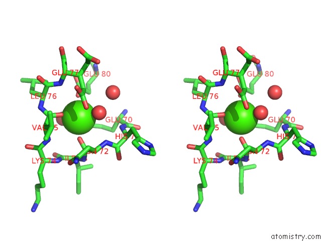
Calcium Binding Sites:
The binding sites of Calcium atom in the Crystal Structure of Mesotrypsin in Complex with Appi-M17C/I18F/F34C
(pdb code 6har). This binding sites where shown within
5.0 Angstroms radius around Calcium atom.
In total only one binding site of Calcium was determined in the Crystal Structure of Mesotrypsin in Complex with Appi-M17C/I18F/F34C, PDB code: 6har:
In total only one binding site of Calcium was determined in the Crystal Structure of Mesotrypsin in Complex with Appi-M17C/I18F/F34C, PDB code: 6har:
Calcium binding site 1 out of 1 in 6har
Go back to
Calcium binding site 1 out
of 1 in the Crystal Structure of Mesotrypsin in Complex with Appi-M17C/I18F/F34C

Mono view
Stereo pair view
Mono view
Stereo pair view
A full contact list of Calcium with other atoms in the Ca binding
site number 1 of Crystal Structure of Mesotrypsin in Complex with Appi-M17C/I18F/F34C within 5.0Å range:
|
Reference:
I.Cohen,
M.Coban,
A.Shahar,
B.Sankaran,
A.Hockla,
S.Lacham,
T.R.Caulfield,
E.S.Radisky,
N.Papo.
Disulfide Engineering of Human Kunitz-Type Serine Protease Inhibitors Enhances Proteolytic Stability and Target Affinity Toward Mesotrypsin. J.Biol.Chem. V. 294 5105 2019.
ISSN: ESSN 1083-351X
PubMed: 30700553
DOI: 10.1074/JBC.RA118.007292
Page generated: Wed Jul 9 14:33:48 2025
ISSN: ESSN 1083-351X
PubMed: 30700553
DOI: 10.1074/JBC.RA118.007292
Last articles
Mg in 6AVTMg in 6AVM
Mg in 6AVP
Mg in 6AUM
Mg in 6ATG
Mg in 6AU9
Mg in 6AT2
Mg in 6AU6
Mg in 6ASW
Mg in 6ASO