Calcium »
PDB 6hi0-6i1q »
6hpe »
Calcium in PDB 6hpe: Crystal Structure of Glutathione Transferase Omega 3S From Trametes Versicolor in Complex with the Glutathione Adduct of Phenethyl- Isothiocyanate
Protein crystallography data
The structure of Crystal Structure of Glutathione Transferase Omega 3S From Trametes Versicolor in Complex with the Glutathione Adduct of Phenethyl- Isothiocyanate, PDB code: 6hpe
was solved by
M.Schwartz,
F.Favier,
C.Didierjean,
with X-Ray Crystallography technique. A brief refinement statistics is given in the table below:
| Resolution Low / High (Å) | 46.74 / 1.45 |
| Space group | P 21 21 21 |
| Cell size a, b, c (Å), α, β, γ (°) | 50.260, 104.030, 106.470, 90.00, 90.00, 90.00 |
| R / Rfree (%) | 16 / 18.2 |
Calcium Binding Sites:
The binding sites of Calcium atom in the Crystal Structure of Glutathione Transferase Omega 3S From Trametes Versicolor in Complex with the Glutathione Adduct of Phenethyl- Isothiocyanate
(pdb code 6hpe). This binding sites where shown within
5.0 Angstroms radius around Calcium atom.
In total only one binding site of Calcium was determined in the Crystal Structure of Glutathione Transferase Omega 3S From Trametes Versicolor in Complex with the Glutathione Adduct of Phenethyl- Isothiocyanate, PDB code: 6hpe:
In total only one binding site of Calcium was determined in the Crystal Structure of Glutathione Transferase Omega 3S From Trametes Versicolor in Complex with the Glutathione Adduct of Phenethyl- Isothiocyanate, PDB code: 6hpe:
Calcium binding site 1 out of 1 in 6hpe
Go back to
Calcium binding site 1 out
of 1 in the Crystal Structure of Glutathione Transferase Omega 3S From Trametes Versicolor in Complex with the Glutathione Adduct of Phenethyl- Isothiocyanate
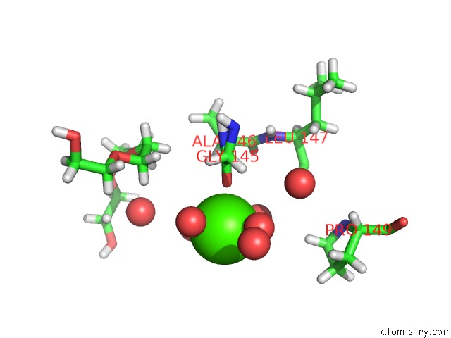
Mono view
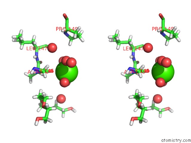
Stereo pair view
Mono view
Stereo pair view
A full contact list of Calcium with other atoms in the Ca binding
site number 1 of Crystal Structure of Glutathione Transferase Omega 3S From Trametes Versicolor in Complex with the Glutathione Adduct of Phenethyl- Isothiocyanate within 5.0Å range:
|
Reference:
M.Schwartz,
T.Perrot,
M.Morel-Rouhier,
G.Mulliert,
E.Gelhaye,
C.Didierjean,
F.Favier.
The Structure of Trametes Versicolor Glutathione Transferase Omega 3S Bound to Its Conjugation Product Glutathionyl-Phenethylthiocarbamate Reveals Plasticity of Its Active Site. Protein Sci. V. 28 1143 2019.
ISSN: ESSN 1469-896X
PubMed: 30972861
DOI: 10.1002/PRO.3620
Page generated: Wed Jul 9 14:40:39 2025
ISSN: ESSN 1469-896X
PubMed: 30972861
DOI: 10.1002/PRO.3620
Last articles
Mg in 2UU7Mg in 2UAG
Mg in 2UKD
Mg in 2SHK
Mg in 2TPS
Mg in 2TRT
Mg in 2TRA
Mg in 2RMK
Mg in 2RUS
Mg in 2TCT