Calcium »
PDB 6hi0-6i1q »
6hqb »
Calcium in PDB 6hqb: Monomeric Cyanobacterial Photosystem I
Enzymatic activity of Monomeric Cyanobacterial Photosystem I
All present enzymatic activity of Monomeric Cyanobacterial Photosystem I:
1.97.1.12;
1.97.1.12;
Protein crystallography data
The structure of Monomeric Cyanobacterial Photosystem I, PDB code: 6hqb
was solved by
S.Y.Netzer-El,
N.Nelson,
I.Caspy,
with X-Ray Crystallography technique. A brief refinement statistics is given in the table below:
| Resolution Low / High (Å) | 49.29 / 4.00 |
| Space group | P 21 21 21 |
| Cell size a, b, c (Å), α, β, γ (°) | 124.322, 178.658, 181.446, 90.00, 90.00, 90.00 |
| R / Rfree (%) | 25.5 / 29.5 |
Other elements in 6hqb:
The structure of Monomeric Cyanobacterial Photosystem I also contains other interesting chemical elements:
| Magnesium | (Mg) | 93 atoms |
| Iron | (Fe) | 12 atoms |
| Chlorine | (Cl) | 1 atom |
Calcium Binding Sites:
The binding sites of Calcium atom in the Monomeric Cyanobacterial Photosystem I
(pdb code 6hqb). This binding sites where shown within
5.0 Angstroms radius around Calcium atom.
In total 2 binding sites of Calcium where determined in the Monomeric Cyanobacterial Photosystem I, PDB code: 6hqb:
Jump to Calcium binding site number: 1; 2;
In total 2 binding sites of Calcium where determined in the Monomeric Cyanobacterial Photosystem I, PDB code: 6hqb:
Jump to Calcium binding site number: 1; 2;
Calcium binding site 1 out of 2 in 6hqb
Go back to
Calcium binding site 1 out
of 2 in the Monomeric Cyanobacterial Photosystem I
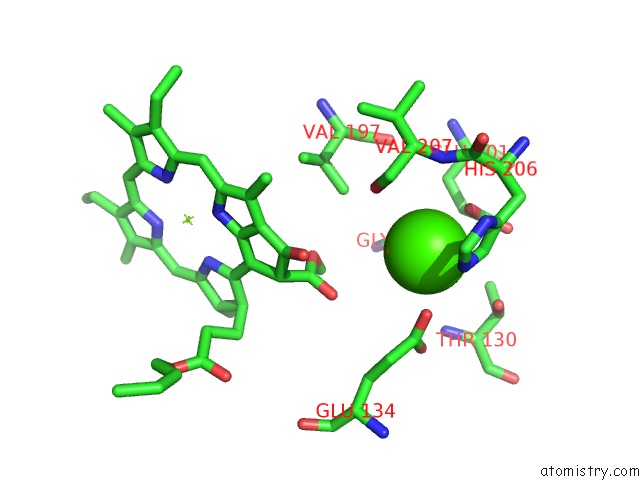
Mono view
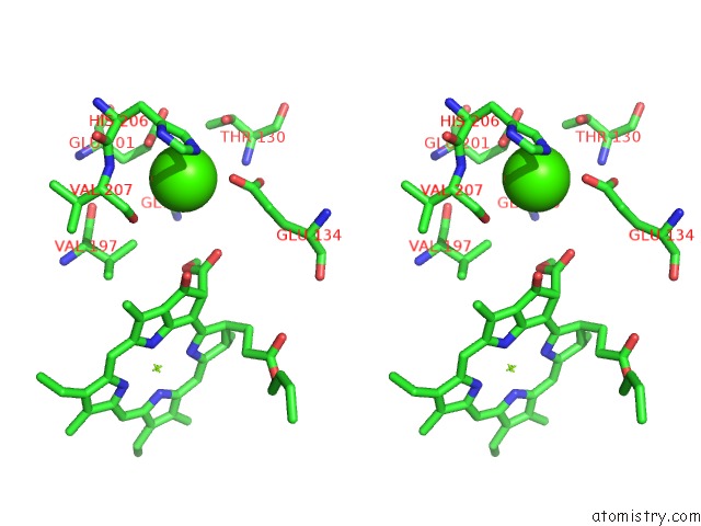
Stereo pair view
Mono view
Stereo pair view
A full contact list of Calcium with other atoms in the Ca binding
site number 1 of Monomeric Cyanobacterial Photosystem I within 5.0Å range:
|
Calcium binding site 2 out of 2 in 6hqb
Go back to
Calcium binding site 2 out
of 2 in the Monomeric Cyanobacterial Photosystem I
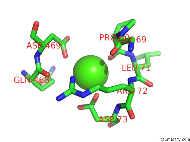
Mono view
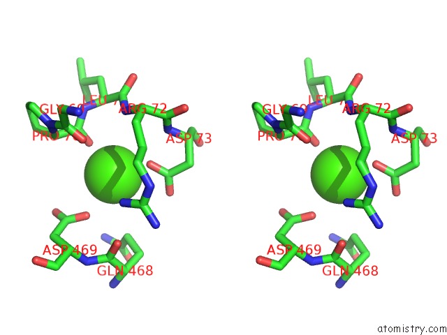
Stereo pair view
Mono view
Stereo pair view
A full contact list of Calcium with other atoms in the Ca binding
site number 2 of Monomeric Cyanobacterial Photosystem I within 5.0Å range:
|
Reference:
S.Y.Netzer-El,
I.Caspy,
N.Nelson.
Crystal Structure of Photosystem I Monomer From Synechocystis Pcc 6803. Front Plant Sci V. 9 1865 2018.
ISSN: ESSN 1664-462X
PubMed: 30662446
DOI: 10.3389/FPLS.2018.01865
Page generated: Wed Jul 9 14:41:57 2025
ISSN: ESSN 1664-462X
PubMed: 30662446
DOI: 10.3389/FPLS.2018.01865
Last articles
K in 9BKDK in 9BWS
K in 9BWR
K in 9BSN
K in 9BNJ
K in 9BWQ
K in 9B76
K in 9B8U
K in 8ZAQ
K in 8ZNT