Calcium »
PDB 6hi0-6i1q »
6hzz »
Calcium in PDB 6hzz: Structure of Human D-Glucuronyl C5 Epimerase
Enzymatic activity of Structure of Human D-Glucuronyl C5 Epimerase
All present enzymatic activity of Structure of Human D-Glucuronyl C5 Epimerase:
5.1.3.17;
5.1.3.17;
Protein crystallography data
The structure of Structure of Human D-Glucuronyl C5 Epimerase, PDB code: 6hzz
was solved by
C.Debarnot,
Y.R.Monneau,
V.Roig-Zamboni,
C.Le Narvor,
A.Goulet,
F.Fadel,
R.R.Vives,
D.Bonnaffe,
H.Lortat-Jacob,
Y.Bourne,
with X-Ray Crystallography technique. A brief refinement statistics is given in the table below:
| Resolution Low / High (Å) | 43.83 / 2.52 |
| Space group | P 61 |
| Cell size a, b, c (Å), α, β, γ (°) | 99.820, 99.820, 262.970, 90.00, 90.00, 120.00 |
| R / Rfree (%) | 17.6 / 22.6 |
Other elements in 6hzz:
The structure of Structure of Human D-Glucuronyl C5 Epimerase also contains other interesting chemical elements:
| Chlorine | (Cl) | 2 atoms |
Calcium Binding Sites:
The binding sites of Calcium atom in the Structure of Human D-Glucuronyl C5 Epimerase
(pdb code 6hzz). This binding sites where shown within
5.0 Angstroms radius around Calcium atom.
In total 2 binding sites of Calcium where determined in the Structure of Human D-Glucuronyl C5 Epimerase, PDB code: 6hzz:
Jump to Calcium binding site number: 1; 2;
In total 2 binding sites of Calcium where determined in the Structure of Human D-Glucuronyl C5 Epimerase, PDB code: 6hzz:
Jump to Calcium binding site number: 1; 2;
Calcium binding site 1 out of 2 in 6hzz
Go back to
Calcium binding site 1 out
of 2 in the Structure of Human D-Glucuronyl C5 Epimerase
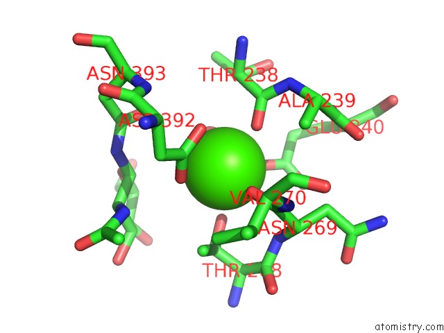
Mono view
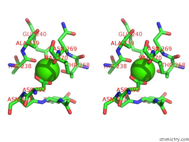
Stereo pair view
Mono view
Stereo pair view
A full contact list of Calcium with other atoms in the Ca binding
site number 1 of Structure of Human D-Glucuronyl C5 Epimerase within 5.0Å range:
|
Calcium binding site 2 out of 2 in 6hzz
Go back to
Calcium binding site 2 out
of 2 in the Structure of Human D-Glucuronyl C5 Epimerase
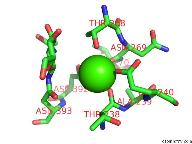
Mono view
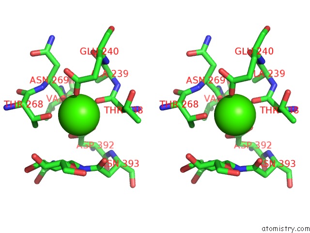
Stereo pair view
Mono view
Stereo pair view
A full contact list of Calcium with other atoms in the Ca binding
site number 2 of Structure of Human D-Glucuronyl C5 Epimerase within 5.0Å range:
|
Reference:
C.Debarnot,
Y.R.Monneau,
V.Roig-Zamboni,
V.Delauzun,
C.Le Narvor,
E.Richard,
J.Henault,
A.Goulet,
F.Fadel,
R.R.Vives,
B.Priem,
D.Bonnaffe,
H.Lortat-Jacob,
Y.Bourne.
Substrate Binding Mode and Catalytic Mechanism of Human Heparan Sulfate D-Glucuronyl C5 Epimerase. Proc.Natl.Acad.Sci.Usa V. 116 6760 2019.
ISSN: ESSN 1091-6490
PubMed: 30872481
DOI: 10.1073/PNAS.1818333116
Page generated: Wed Jul 9 14:45:25 2025
ISSN: ESSN 1091-6490
PubMed: 30872481
DOI: 10.1073/PNAS.1818333116
Last articles
Mg in 3A16Mg in 3A1C
Mg in 3A14
Mg in 3A11
Mg in 3A10
Mg in 3A0U
Mg in 3A0H
Mg in 3A0B
Mg in 364D
Mg in 3A0T