Calcium »
PDB 6i1t-6im1 »
6i28 »
Calcium in PDB 6i28: Crystal Structure of Cydia Pomonella Ptp-2 Phosphatase
Protein crystallography data
The structure of Crystal Structure of Cydia Pomonella Ptp-2 Phosphatase, PDB code: 6i28
was solved by
G.Huang,
J.P.Keown,
M.R.Oliver,
P.Metcalf,
with X-Ray Crystallography technique. A brief refinement statistics is given in the table below:
| Resolution Low / High (Å) | 33.70 / 1.65 |
| Space group | I 2 2 2 |
| Cell size a, b, c (Å), α, β, γ (°) | 64.690, 66.720, 97.960, 90.00, 90.00, 90.00 |
| R / Rfree (%) | 18.5 / 22.4 |
Calcium Binding Sites:
The binding sites of Calcium atom in the Crystal Structure of Cydia Pomonella Ptp-2 Phosphatase
(pdb code 6i28). This binding sites where shown within
5.0 Angstroms radius around Calcium atom.
In total only one binding site of Calcium was determined in the Crystal Structure of Cydia Pomonella Ptp-2 Phosphatase, PDB code: 6i28:
In total only one binding site of Calcium was determined in the Crystal Structure of Cydia Pomonella Ptp-2 Phosphatase, PDB code: 6i28:
Calcium binding site 1 out of 1 in 6i28
Go back to
Calcium binding site 1 out
of 1 in the Crystal Structure of Cydia Pomonella Ptp-2 Phosphatase

Mono view
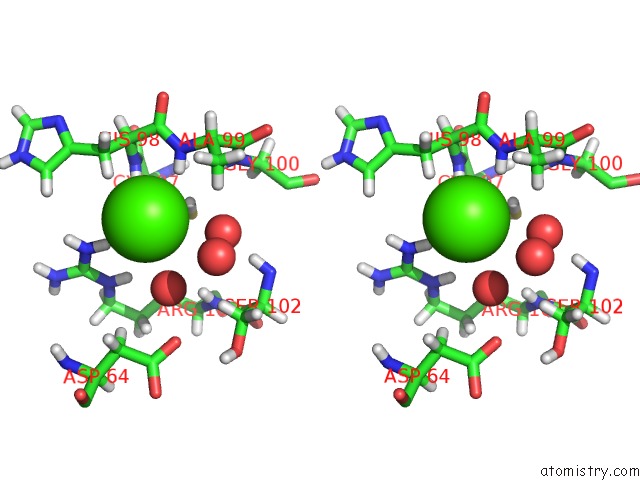
Stereo pair view
Mono view
Stereo pair view
A full contact list of Calcium with other atoms in the Ca binding
site number 1 of Crystal Structure of Cydia Pomonella Ptp-2 Phosphatase within 5.0Å range:
|
Reference:
G.Huang,
M.R.Oliver,
J.R.Keown,
D.C.Goldstone,
P.Metcalf.
Crystal Structure of Protein Tyrosine Phosphatase-2 From Cydia Pomonella Granulovirus. Acta Crystallogr.,Sect.F V. 75 233 2019.
ISSN: ESSN 2053-230X
PubMed: 30950823
DOI: 10.1107/S2053230X19002322
Page generated: Wed Jul 9 14:46:10 2025
ISSN: ESSN 2053-230X
PubMed: 30950823
DOI: 10.1107/S2053230X19002322
Last articles
Mg in 4DV0Mg in 4DV1
Mg in 4DUZ
Mg in 4DUY
Mg in 4DR7
Mg in 4DR6
Mg in 4DR5
Mg in 4DUX
Mg in 4DUW
Mg in 4DUV