Calcium »
PDB 6im3-6j43 »
6iqq »
Calcium in PDB 6iqq: Crystal Structure of Prc with S452I and L252Y Mutations in Complex with Nlpi
Enzymatic activity of Crystal Structure of Prc with S452I and L252Y Mutations in Complex with Nlpi
All present enzymatic activity of Crystal Structure of Prc with S452I and L252Y Mutations in Complex with Nlpi:
3.4.21.102;
3.4.21.102;
Protein crystallography data
The structure of Crystal Structure of Prc with S452I and L252Y Mutations in Complex with Nlpi, PDB code: 6iqq
was solved by
C.K.Chueh,
C.I.Chang,
with X-Ray Crystallography technique. A brief refinement statistics is given in the table below:
| Resolution Low / High (Å) | 29.98 / 2.80 |
| Space group | P 21 21 21 |
| Cell size a, b, c (Å), α, β, γ (°) | 118.936, 143.882, 153.599, 90.00, 90.00, 90.00 |
| R / Rfree (%) | 22.2 / 27.8 |
Calcium Binding Sites:
The binding sites of Calcium atom in the Crystal Structure of Prc with S452I and L252Y Mutations in Complex with Nlpi
(pdb code 6iqq). This binding sites where shown within
5.0 Angstroms radius around Calcium atom.
In total 2 binding sites of Calcium where determined in the Crystal Structure of Prc with S452I and L252Y Mutations in Complex with Nlpi, PDB code: 6iqq:
Jump to Calcium binding site number: 1; 2;
In total 2 binding sites of Calcium where determined in the Crystal Structure of Prc with S452I and L252Y Mutations in Complex with Nlpi, PDB code: 6iqq:
Jump to Calcium binding site number: 1; 2;
Calcium binding site 1 out of 2 in 6iqq
Go back to
Calcium binding site 1 out
of 2 in the Crystal Structure of Prc with S452I and L252Y Mutations in Complex with Nlpi
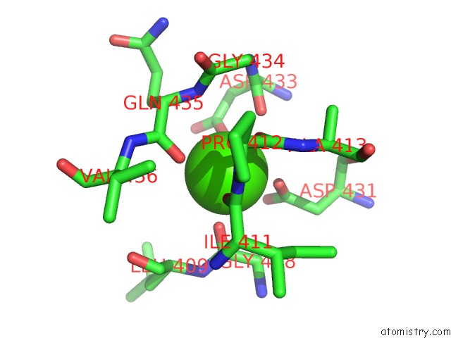
Mono view
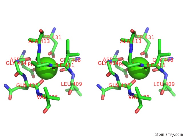
Stereo pair view
Mono view
Stereo pair view
A full contact list of Calcium with other atoms in the Ca binding
site number 1 of Crystal Structure of Prc with S452I and L252Y Mutations in Complex with Nlpi within 5.0Å range:
|
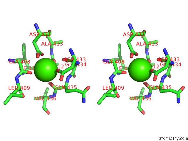
Calcium binding site 2 out of 2 in 6iqq
Go back to
Calcium binding site 2 out
of 2 in the Crystal Structure of Prc with S452I and L252Y Mutations in Complex with Nlpi
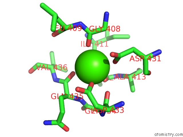
Mono view
Stereo pair view
Mono view
Stereo pair view
A full contact list of Calcium with other atoms in the Ca binding
site number 2 of Crystal Structure of Prc with S452I and L252Y Mutations in Complex with Nlpi within 5.0Å range:
|
Reference:
C.K.Chueh,
N.Som,
L.C.Ke,
M.R.Ho,
M.Reddy,
C.I.Chang.
Structural Basis For the Differential Regulatory Roles of the Pdz Domain in C-Terminal Processing Proteases. Mbio V. 10 2019.
ISSN: ESSN 2150-7511
PubMed: 31387902
DOI: 10.1128/MBIO.01129-19
Page generated: Wed Jul 9 14:58:30 2025
ISSN: ESSN 2150-7511
PubMed: 31387902
DOI: 10.1128/MBIO.01129-19
Last articles
Fe in 2YXOFe in 2YRS
Fe in 2YXC
Fe in 2YNM
Fe in 2YVJ
Fe in 2YP1
Fe in 2YU2
Fe in 2YU1
Fe in 2YQB
Fe in 2YOO