Calcium »
PDB 6j4h-6jlm »
6jhf »
Calcium in PDB 6jhf: Crystal Structure of Apo Pullulanase From Paenibacillus Barengoltzii
Enzymatic activity of Crystal Structure of Apo Pullulanase From Paenibacillus Barengoltzii
All present enzymatic activity of Crystal Structure of Apo Pullulanase From Paenibacillus Barengoltzii:
3.2.1.41;
3.2.1.41;
Protein crystallography data
The structure of Crystal Structure of Apo Pullulanase From Paenibacillus Barengoltzii, PDB code: 6jhf
was solved by
S.W.Wu,
S.Q.Yang,
Z.Qin,
X.You,
P.Huang,
Z.Q.Jiang,
with X-Ray Crystallography technique. A brief refinement statistics is given in the table below:
| Resolution Low / High (Å) | 49.29 / 1.71 |
| Space group | P 2 21 21 |
| Cell size a, b, c (Å), α, β, γ (°) | 50.395, 98.583, 142.019, 90.00, 90.00, 90.00 |
| R / Rfree (%) | 15.2 / 18.2 |
Other elements in 6jhf:
The structure of Crystal Structure of Apo Pullulanase From Paenibacillus Barengoltzii also contains other interesting chemical elements:
| Chlorine | (Cl) | 2 atoms |
Calcium Binding Sites:
The binding sites of Calcium atom in the Crystal Structure of Apo Pullulanase From Paenibacillus Barengoltzii
(pdb code 6jhf). This binding sites where shown within
5.0 Angstroms radius around Calcium atom.
In total only one binding site of Calcium was determined in the Crystal Structure of Apo Pullulanase From Paenibacillus Barengoltzii, PDB code: 6jhf:
In total only one binding site of Calcium was determined in the Crystal Structure of Apo Pullulanase From Paenibacillus Barengoltzii, PDB code: 6jhf:
Calcium binding site 1 out of 1 in 6jhf
Go back to
Calcium binding site 1 out
of 1 in the Crystal Structure of Apo Pullulanase From Paenibacillus Barengoltzii
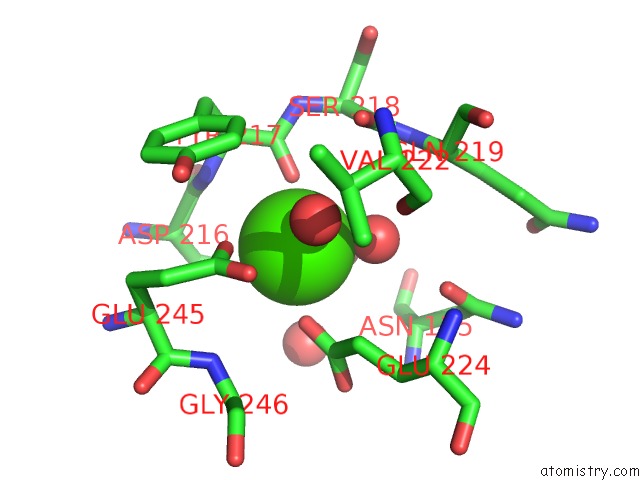
Mono view
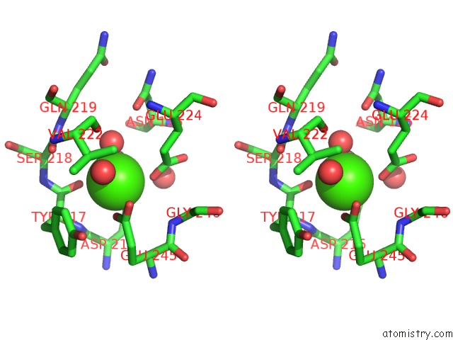
Stereo pair view
Mono view
Stereo pair view
A full contact list of Calcium with other atoms in the Ca binding
site number 1 of Crystal Structure of Apo Pullulanase From Paenibacillus Barengoltzii within 5.0Å range:
|
Reference:
S.W.Wu,
S.Q.Yang,
Z.Qin,
X.You,
P.Huang,
Z.Q.Jiang.
Crystal Structure of Apo Pullulanase From Paenibacillus Barengoltzii To Be Published.
Page generated: Wed Jul 9 15:07:21 2025
Last articles
Mo in 1DGJMo in 1DMR
Mo in 1AA6
Mn in 9ONQ
Mn in 9XIM
Mn in 9XIA
Mn in 9NJO
Mn in 9QB8
Mn in 9MR6
Mn in 9M2Q