Calcium »
PDB 6jln-6k2p »
6jwt »
Calcium in PDB 6jwt: Crystal Structure of Plasmodium Falciparum Hppk-Dhps S436F/A437G/A613T Triple Mutant with Pteroate
Protein crystallography data
The structure of Crystal Structure of Plasmodium Falciparum Hppk-Dhps S436F/A437G/A613T Triple Mutant with Pteroate, PDB code: 6jwt
was solved by
P.Chitnumsub,
A.Jaruwat,
Y.Yuthavong,
with X-Ray Crystallography technique. A brief refinement statistics is given in the table below:
| Resolution Low / High (Å) | 47.26 / 2.56 |
| Space group | P 21 21 21 |
| Cell size a, b, c (Å), α, β, γ (°) | 100.627, 136.597, 138.518, 90.00, 90.00, 90.00 |
| R / Rfree (%) | 19.6 / 24.3 |
Other elements in 6jwt:
The structure of Crystal Structure of Plasmodium Falciparum Hppk-Dhps S436F/A437G/A613T Triple Mutant with Pteroate also contains other interesting chemical elements:
| Magnesium | (Mg) | 3 atoms |
Calcium Binding Sites:
The binding sites of Calcium atom in the Crystal Structure of Plasmodium Falciparum Hppk-Dhps S436F/A437G/A613T Triple Mutant with Pteroate
(pdb code 6jwt). This binding sites where shown within
5.0 Angstroms radius around Calcium atom.
In total 2 binding sites of Calcium where determined in the Crystal Structure of Plasmodium Falciparum Hppk-Dhps S436F/A437G/A613T Triple Mutant with Pteroate, PDB code: 6jwt:
Jump to Calcium binding site number: 1; 2;
In total 2 binding sites of Calcium where determined in the Crystal Structure of Plasmodium Falciparum Hppk-Dhps S436F/A437G/A613T Triple Mutant with Pteroate, PDB code: 6jwt:
Jump to Calcium binding site number: 1; 2;
Calcium binding site 1 out of 2 in 6jwt
Go back to
Calcium binding site 1 out
of 2 in the Crystal Structure of Plasmodium Falciparum Hppk-Dhps S436F/A437G/A613T Triple Mutant with Pteroate
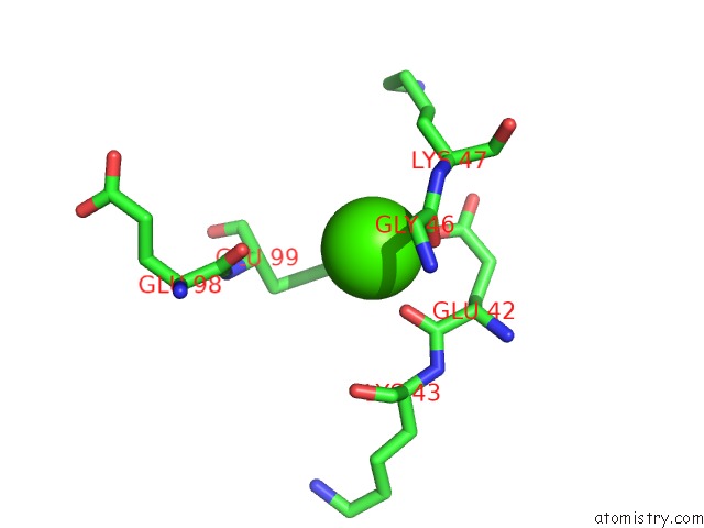
Mono view
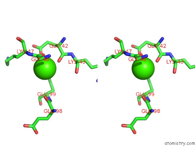
Stereo pair view
Mono view
Stereo pair view
A full contact list of Calcium with other atoms in the Ca binding
site number 1 of Crystal Structure of Plasmodium Falciparum Hppk-Dhps S436F/A437G/A613T Triple Mutant with Pteroate within 5.0Å range:
|
Calcium binding site 2 out of 2 in 6jwt
Go back to
Calcium binding site 2 out
of 2 in the Crystal Structure of Plasmodium Falciparum Hppk-Dhps S436F/A437G/A613T Triple Mutant with Pteroate
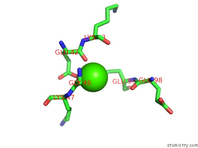
Mono view
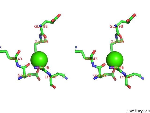
Stereo pair view
Mono view
Stereo pair view
A full contact list of Calcium with other atoms in the Ca binding
site number 2 of Crystal Structure of Plasmodium Falciparum Hppk-Dhps S436F/A437G/A613T Triple Mutant with Pteroate within 5.0Å range:
|
Reference:
P.Chitnumsub,
A.Jaruwat,
Y.Talawanich,
K.Noytanom,
B.Liwnaree,
S.Poen,
Y.Yuthavong.
The Structure of Plasmodium Falciparum Hydroxymethyldihydropterin Pyrophosphokinase-Dihydropteroate Synthase Reveals the Basis of Sulfa Resistance. Febs J. 2019.
ISSN: ISSN 1742-464X
PubMed: 31883412
DOI: 10.1111/FEBS.15196
Page generated: Wed Jul 9 15:20:28 2025
ISSN: ISSN 1742-464X
PubMed: 31883412
DOI: 10.1111/FEBS.15196
Last articles
Mg in 4X5CMg in 4X5E
Mg in 4X5V
Mg in 4X5B
Mg in 4X59
Mg in 4X58
Mg in 4X4V
Mg in 4X4S
Mg in 4X4R
Mg in 4X4Q