Calcium »
PDB 6kg9-6kzk »
6khx »
Calcium in PDB 6khx: Crystal Structure of Prx From Akkermansia Muciniphila
Protein crystallography data
The structure of Crystal Structure of Prx From Akkermansia Muciniphila, PDB code: 6khx
was solved by
M.Li,
J.Wang,
W.Xu,
Y.Wang,
M.Zhang,
M.Wang,
with X-Ray Crystallography technique. A brief refinement statistics is given in the table below:
| Resolution Low / High (Å) | 49.10 / 2.58 |
| Space group | P 21 21 2 |
| Cell size a, b, c (Å), α, β, γ (°) | 132.733, 211.371, 92.802, 90.00, 90.00, 90.00 |
| R / Rfree (%) | 17.7 / 21.8 |
Calcium Binding Sites:
The binding sites of Calcium atom in the Crystal Structure of Prx From Akkermansia Muciniphila
(pdb code 6khx). This binding sites where shown within
5.0 Angstroms radius around Calcium atom.
In total 8 binding sites of Calcium where determined in the Crystal Structure of Prx From Akkermansia Muciniphila, PDB code: 6khx:
Jump to Calcium binding site number: 1; 2; 3; 4; 5; 6; 7; 8;
In total 8 binding sites of Calcium where determined in the Crystal Structure of Prx From Akkermansia Muciniphila, PDB code: 6khx:
Jump to Calcium binding site number: 1; 2; 3; 4; 5; 6; 7; 8;
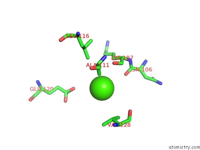
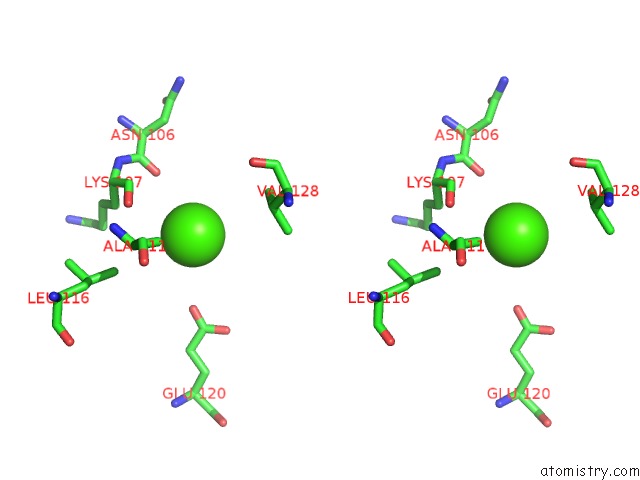
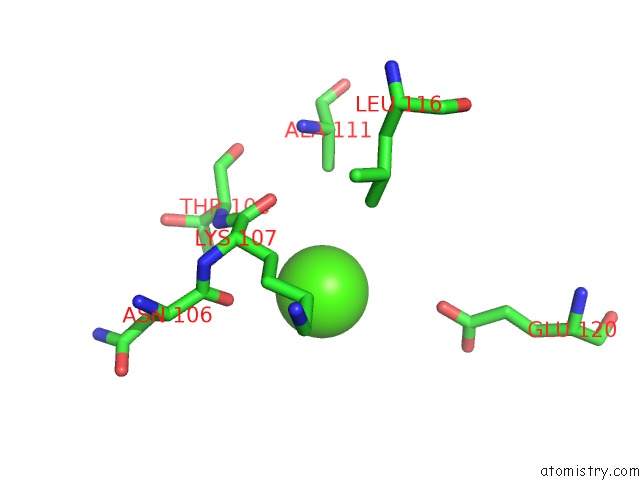
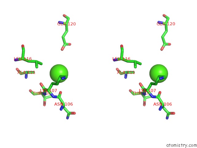
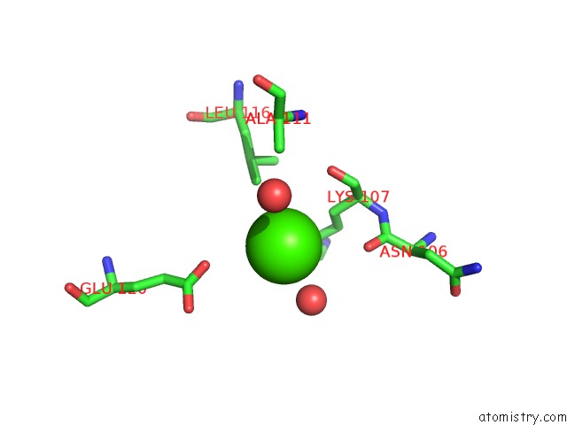
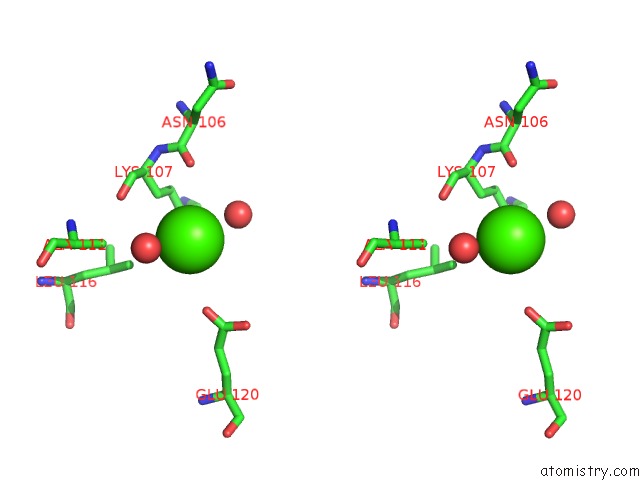
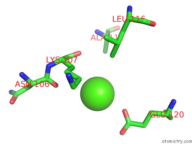
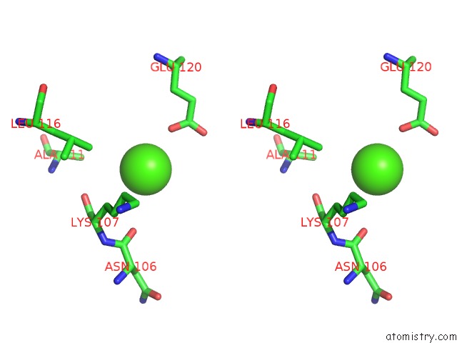
Calcium binding site 1 out of 8 in 6khx
Go back to
Calcium binding site 1 out
of 8 in the Crystal Structure of Prx From Akkermansia Muciniphila
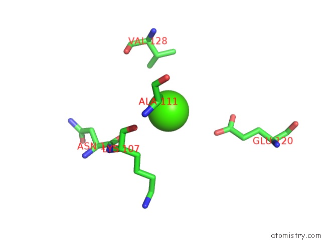
Mono view
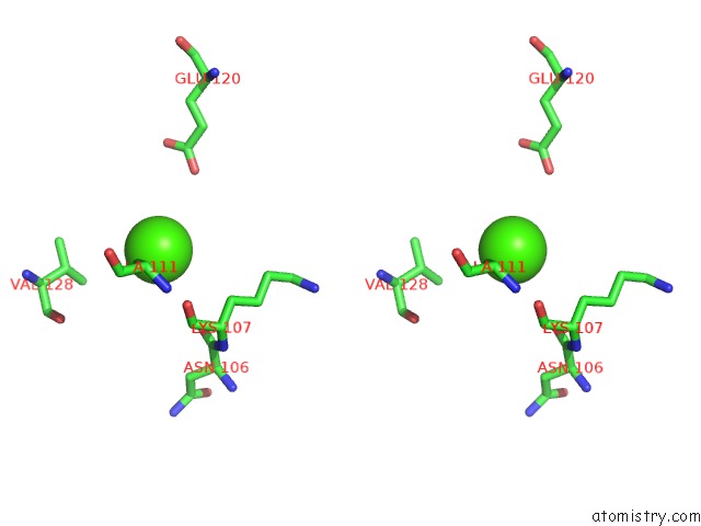
Stereo pair view
Mono view
Stereo pair view
A full contact list of Calcium with other atoms in the Ca binding
site number 1 of Crystal Structure of Prx From Akkermansia Muciniphila within 5.0Å range:
|
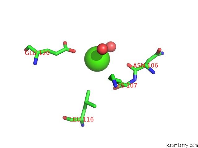
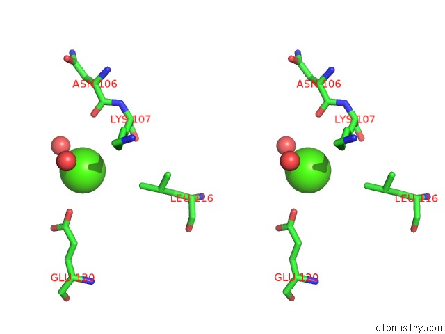
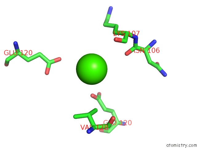
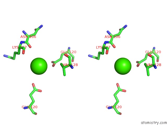
Calcium binding site 2 out of 8 in 6khx
Go back to
Calcium binding site 2 out
of 8 in the Crystal Structure of Prx From Akkermansia Muciniphila
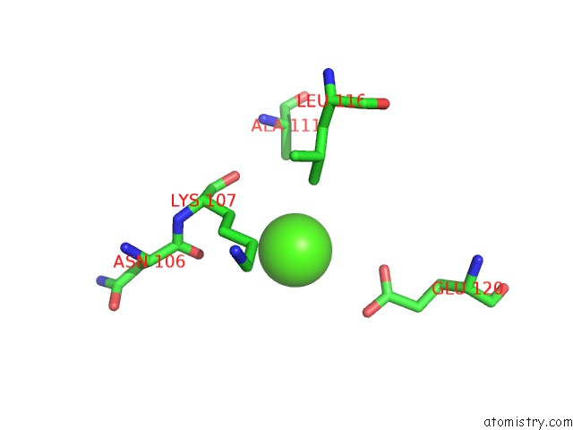
Mono view
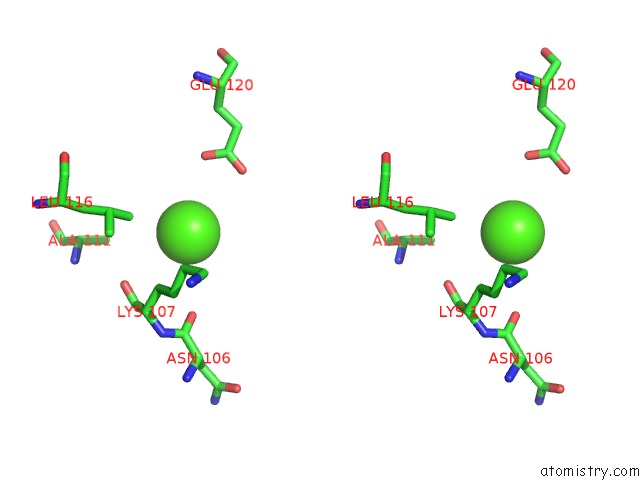
Stereo pair view
Mono view
Stereo pair view
A full contact list of Calcium with other atoms in the Ca binding
site number 2 of Crystal Structure of Prx From Akkermansia Muciniphila within 5.0Å range:
|
Calcium binding site 3 out of 8 in 6khx
Go back to
Calcium binding site 3 out
of 8 in the Crystal Structure of Prx From Akkermansia Muciniphila
Mono view
Stereo pair view
Mono view
Stereo pair view
A full contact list of Calcium with other atoms in the Ca binding
site number 3 of Crystal Structure of Prx From Akkermansia Muciniphila within 5.0Å range:
|
Calcium binding site 4 out of 8 in 6khx
Go back to
Calcium binding site 4 out
of 8 in the Crystal Structure of Prx From Akkermansia Muciniphila
Mono view
Stereo pair view
Mono view
Stereo pair view
A full contact list of Calcium with other atoms in the Ca binding
site number 4 of Crystal Structure of Prx From Akkermansia Muciniphila within 5.0Å range:
|
Calcium binding site 5 out of 8 in 6khx
Go back to
Calcium binding site 5 out
of 8 in the Crystal Structure of Prx From Akkermansia Muciniphila
Mono view
Stereo pair view
Mono view
Stereo pair view
A full contact list of Calcium with other atoms in the Ca binding
site number 5 of Crystal Structure of Prx From Akkermansia Muciniphila within 5.0Å range:
|
Calcium binding site 6 out of 8 in 6khx
Go back to
Calcium binding site 6 out
of 8 in the Crystal Structure of Prx From Akkermansia Muciniphila
Mono view
Stereo pair view
Mono view
Stereo pair view
A full contact list of Calcium with other atoms in the Ca binding
site number 6 of Crystal Structure of Prx From Akkermansia Muciniphila within 5.0Å range:
|
Calcium binding site 7 out of 8 in 6khx
Go back to
Calcium binding site 7 out
of 8 in the Crystal Structure of Prx From Akkermansia Muciniphila
Mono view
Stereo pair view
Mono view
Stereo pair view
A full contact list of Calcium with other atoms in the Ca binding
site number 7 of Crystal Structure of Prx From Akkermansia Muciniphila within 5.0Å range:
|
Calcium binding site 8 out of 8 in 6khx
Go back to
Calcium binding site 8 out
of 8 in the Crystal Structure of Prx From Akkermansia Muciniphila
Mono view
Stereo pair view
Mono view
Stereo pair view
A full contact list of Calcium with other atoms in the Ca binding
site number 8 of Crystal Structure of Prx From Akkermansia Muciniphila within 5.0Å range:
|
Reference:
M.Li,
J.Wang,
W.Xu,
Y.Wang,
M.Zhang,
M.Wang.
Crystal Structure of Akkermansia Muciniphila Peroxiredoxin Reveals A Novel Regulatory Mechanism of Typical 2-Cys Prxs By A Distinct Loop. Febs Lett. 2020.
ISSN: ISSN 0014-5793
PubMed: 32027024
DOI: 10.1002/1873-3468.13753
Page generated: Wed Jul 9 15:34:18 2025
ISSN: ISSN 0014-5793
PubMed: 32027024
DOI: 10.1002/1873-3468.13753
Last articles
Mg in 5N9XMg in 5MX2
Mg in 5N78
Mg in 5N77
Mg in 5N6Y
Mg in 5N75
Mg in 5N6O
Mg in 5N5N
Mg in 5N69
Mg in 5N6A