Calcium »
PDB 6kg9-6kzk »
6kma »
Calcium in PDB 6kma: Crystal Structure of Suca with Glycolaldehyde-1-13C From Vibrio Vulnificus
Enzymatic activity of Crystal Structure of Suca with Glycolaldehyde-1-13C From Vibrio Vulnificus
All present enzymatic activity of Crystal Structure of Suca with Glycolaldehyde-1-13C From Vibrio Vulnificus:
1.2.4.2;
1.2.4.2;
Protein crystallography data
The structure of Crystal Structure of Suca with Glycolaldehyde-1-13C From Vibrio Vulnificus, PDB code: 6kma
was solved by
P.W.Seo,
J.S.Kim,
with X-Ray Crystallography technique. A brief refinement statistics is given in the table below:
| Resolution Low / High (Å) | 47.65 / 2.28 |
| Space group | P 1 |
| Cell size a, b, c (Å), α, β, γ (°) | 76.820, 84.559, 145.218, 79.51, 88.47, 89.92 |
| R / Rfree (%) | 20 / 23 |
Other elements in 6kma:
The structure of Crystal Structure of Suca with Glycolaldehyde-1-13C From Vibrio Vulnificus also contains other interesting chemical elements:
| Magnesium | (Mg) | 4 atoms |
Calcium Binding Sites:
The binding sites of Calcium atom in the Crystal Structure of Suca with Glycolaldehyde-1-13C From Vibrio Vulnificus
(pdb code 6kma). This binding sites where shown within
5.0 Angstroms radius around Calcium atom.
In total 2 binding sites of Calcium where determined in the Crystal Structure of Suca with Glycolaldehyde-1-13C From Vibrio Vulnificus, PDB code: 6kma:
Jump to Calcium binding site number: 1; 2;
In total 2 binding sites of Calcium where determined in the Crystal Structure of Suca with Glycolaldehyde-1-13C From Vibrio Vulnificus, PDB code: 6kma:
Jump to Calcium binding site number: 1; 2;
Calcium binding site 1 out of 2 in 6kma
Go back to
Calcium binding site 1 out
of 2 in the Crystal Structure of Suca with Glycolaldehyde-1-13C From Vibrio Vulnificus
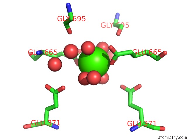
Mono view
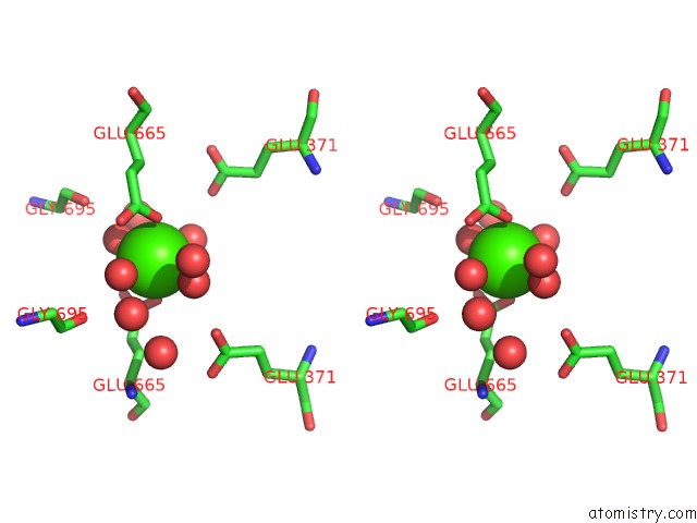
Stereo pair view
Mono view
Stereo pair view
A full contact list of Calcium with other atoms in the Ca binding
site number 1 of Crystal Structure of Suca with Glycolaldehyde-1-13C From Vibrio Vulnificus within 5.0Å range:
|
Calcium binding site 2 out of 2 in 6kma
Go back to
Calcium binding site 2 out
of 2 in the Crystal Structure of Suca with Glycolaldehyde-1-13C From Vibrio Vulnificus
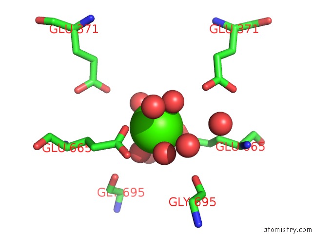
Mono view
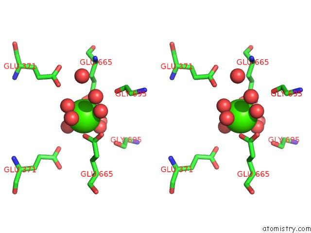
Stereo pair view
Mono view
Stereo pair view
A full contact list of Calcium with other atoms in the Ca binding
site number 2 of Crystal Structure of Suca with Glycolaldehyde-1-13C From Vibrio Vulnificus within 5.0Å range:
|
Reference:
P.W.Seo,
J.S.Kim.
Crystal Structure of Suca with Glycolaldehyde-1-13C From Vibrio Vulnificus at 2.28 Angstroms Resolution. To Be Published.
Page generated: Wed Jul 9 15:36:56 2025
Last articles
Mg in 4LF4Mg in 4LF5
Mg in 4LCZ
Mg in 4LF2
Mg in 4LF1
Mg in 4LEM
Mg in 4LCK
Mg in 4LE0
Mg in 4LDZ
Mg in 4LDT