Calcium »
PDB 6kzo-6ljf »
6l2j »
Calcium in PDB 6l2j: Crystal Structure of Yak Lactoperoxidase at 1.93 A Resolution.
Enzymatic activity of Crystal Structure of Yak Lactoperoxidase at 1.93 A Resolution.
All present enzymatic activity of Crystal Structure of Yak Lactoperoxidase at 1.93 A Resolution.:
1.11.1.7;
1.11.1.7;
Protein crystallography data
The structure of Crystal Structure of Yak Lactoperoxidase at 1.93 A Resolution., PDB code: 6l2j
was solved by
V.Viswanathan,
P.Sharma,
C.Rani,
S.Sharma,
T.P.Singh,
with X-Ray Crystallography technique. A brief refinement statistics is given in the table below:
| Resolution Low / High (Å) | 53.84 / 1.93 |
| Space group | P 1 21 1 |
| Cell size a, b, c (Å), α, β, γ (°) | 53.850, 78.920, 67.750, 90.00, 92.95, 90.00 |
| R / Rfree (%) | 14.5 / 19.8 |
Other elements in 6l2j:
The structure of Crystal Structure of Yak Lactoperoxidase at 1.93 A Resolution. also contains other interesting chemical elements:
| Iodine | (I) | 26 atoms |
| Iron | (Fe) | 1 atom |
Calcium Binding Sites:
The binding sites of Calcium atom in the Crystal Structure of Yak Lactoperoxidase at 1.93 A Resolution.
(pdb code 6l2j). This binding sites where shown within
5.0 Angstroms radius around Calcium atom.
In total 5 binding sites of Calcium where determined in the Crystal Structure of Yak Lactoperoxidase at 1.93 A Resolution., PDB code: 6l2j:
Jump to Calcium binding site number: 1; 2; 3; 4; 5;
In total 5 binding sites of Calcium where determined in the Crystal Structure of Yak Lactoperoxidase at 1.93 A Resolution., PDB code: 6l2j:
Jump to Calcium binding site number: 1; 2; 3; 4; 5;
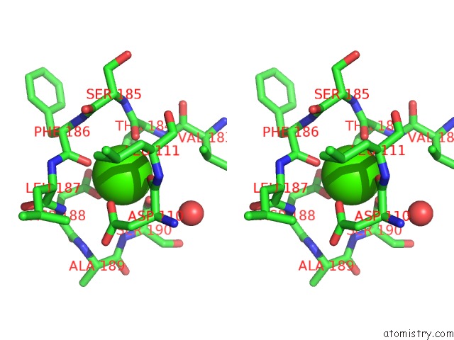
Calcium binding site 1 out of 5 in 6l2j
Go back to
Calcium binding site 1 out
of 5 in the Crystal Structure of Yak Lactoperoxidase at 1.93 A Resolution.
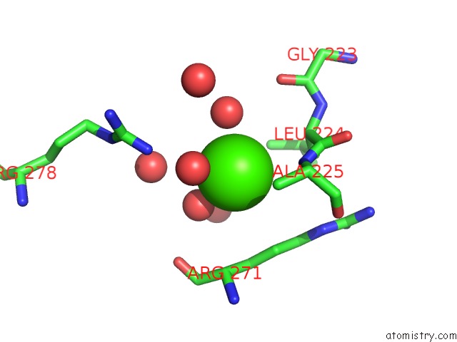
Mono view
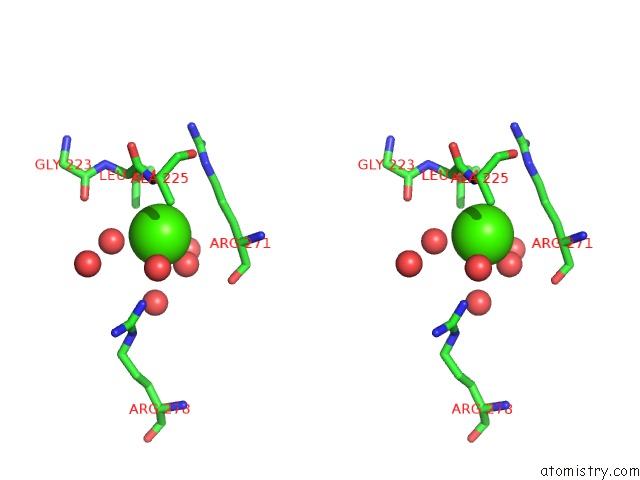
Stereo pair view
Mono view
Stereo pair view
A full contact list of Calcium with other atoms in the Ca binding
site number 1 of Crystal Structure of Yak Lactoperoxidase at 1.93 A Resolution. within 5.0Å range:
|
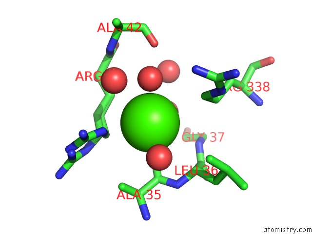
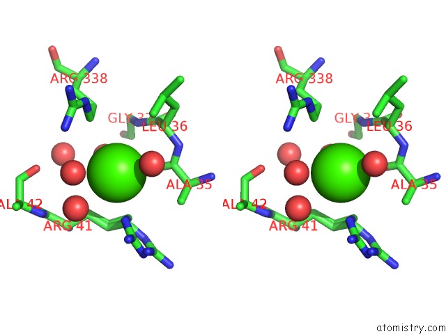
Calcium binding site 2 out of 5 in 6l2j
Go back to
Calcium binding site 2 out
of 5 in the Crystal Structure of Yak Lactoperoxidase at 1.93 A Resolution.
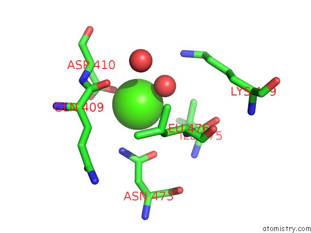
Mono view
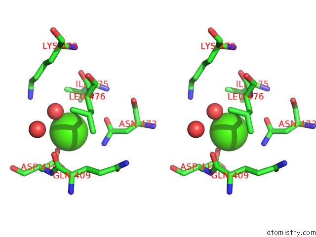
Stereo pair view
Mono view
Stereo pair view
A full contact list of Calcium with other atoms in the Ca binding
site number 2 of Crystal Structure of Yak Lactoperoxidase at 1.93 A Resolution. within 5.0Å range:
|
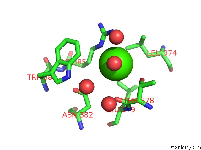
Calcium binding site 3 out of 5 in 6l2j
Go back to
Calcium binding site 3 out
of 5 in the Crystal Structure of Yak Lactoperoxidase at 1.93 A Resolution.

Mono view
Stereo pair view
Mono view
Stereo pair view
A full contact list of Calcium with other atoms in the Ca binding
site number 3 of Crystal Structure of Yak Lactoperoxidase at 1.93 A Resolution. within 5.0Å range:
|
Calcium binding site 4 out of 5 in 6l2j
Go back to
Calcium binding site 4 out
of 5 in the Crystal Structure of Yak Lactoperoxidase at 1.93 A Resolution.
Mono view
Stereo pair view
Mono view
Stereo pair view
A full contact list of Calcium with other atoms in the Ca binding
site number 4 of Crystal Structure of Yak Lactoperoxidase at 1.93 A Resolution. within 5.0Å range:
|
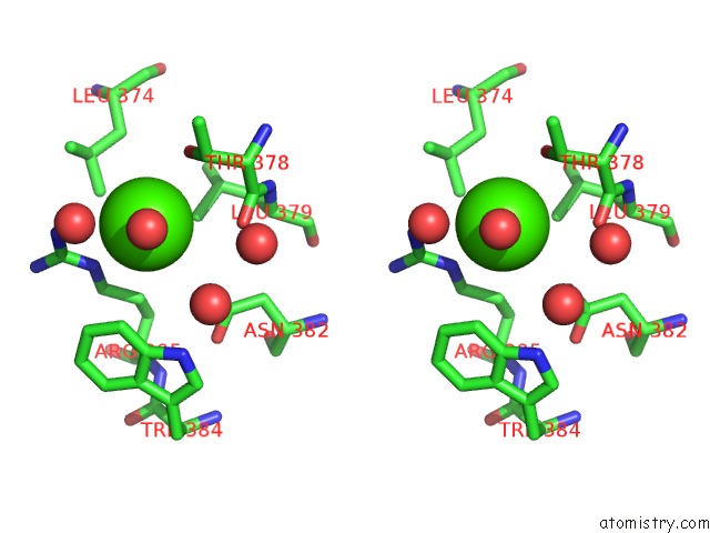
Calcium binding site 5 out of 5 in 6l2j
Go back to
Calcium binding site 5 out
of 5 in the Crystal Structure of Yak Lactoperoxidase at 1.93 A Resolution.
Mono view
Stereo pair view
Mono view
Stereo pair view
A full contact list of Calcium with other atoms in the Ca binding
site number 5 of Crystal Structure of Yak Lactoperoxidase at 1.93 A Resolution. within 5.0Å range:
|
Reference:
V.Viswanathan,
P.Sharma,
C.Rani,
S.Sharma,
T.P.Singh.
Crystal Structure of Yak Lactoperoxidase at 1.93 A Resolution. To Be Published.
Page generated: Wed Jul 9 15:43:23 2025
Last articles
K in 8CGUK in 8CGR
K in 8CGJ
K in 8CGI
K in 8CG1
K in 8CFY
K in 8CG2
K in 8CFX
K in 8CG0
K in 8CFW