Calcium »
PDB 6ljl-6m4s »
6lkc »
Calcium in PDB 6lkc: Crystal Structure of Pfad From Shewanella Piezotolerans in Complex with Fmn
Enzymatic activity of Crystal Structure of Pfad From Shewanella Piezotolerans in Complex with Fmn
All present enzymatic activity of Crystal Structure of Pfad From Shewanella Piezotolerans in Complex with Fmn:
1.3.1.9;
1.3.1.9;
Protein crystallography data
The structure of Crystal Structure of Pfad From Shewanella Piezotolerans in Complex with Fmn, PDB code: 6lkc
was solved by
M.L.Zhang,
Q.Li,
S.S.Meng,
L.J.Guo,
L.He,
J.Z.Huang,
L.Li,
H.D.Zhang,
with X-Ray Crystallography technique. A brief refinement statistics is given in the table below:
| Resolution Low / High (Å) | 53.61 / 2.00 |
| Space group | P 1 21 1 |
| Cell size a, b, c (Å), α, β, γ (°) | 70.467, 62.568, 127.507, 90.00, 105.61, 90.00 |
| R / Rfree (%) | 17.9 / 21.9 |
Calcium Binding Sites:
The binding sites of Calcium atom in the Crystal Structure of Pfad From Shewanella Piezotolerans in Complex with Fmn
(pdb code 6lkc). This binding sites where shown within
5.0 Angstroms radius around Calcium atom.
In total 2 binding sites of Calcium where determined in the Crystal Structure of Pfad From Shewanella Piezotolerans in Complex with Fmn, PDB code: 6lkc:
Jump to Calcium binding site number: 1; 2;
In total 2 binding sites of Calcium where determined in the Crystal Structure of Pfad From Shewanella Piezotolerans in Complex with Fmn, PDB code: 6lkc:
Jump to Calcium binding site number: 1; 2;
Calcium binding site 1 out of 2 in 6lkc
Go back to
Calcium binding site 1 out
of 2 in the Crystal Structure of Pfad From Shewanella Piezotolerans in Complex with Fmn
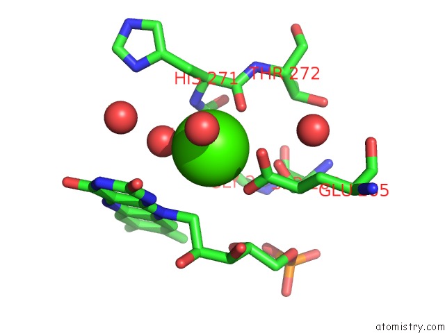
Mono view
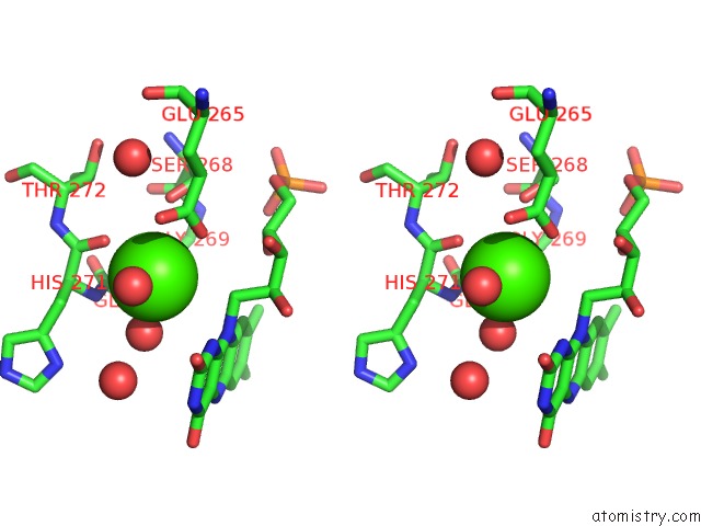
Stereo pair view
Mono view
Stereo pair view
A full contact list of Calcium with other atoms in the Ca binding
site number 1 of Crystal Structure of Pfad From Shewanella Piezotolerans in Complex with Fmn within 5.0Å range:
|
Calcium binding site 2 out of 2 in 6lkc
Go back to
Calcium binding site 2 out
of 2 in the Crystal Structure of Pfad From Shewanella Piezotolerans in Complex with Fmn
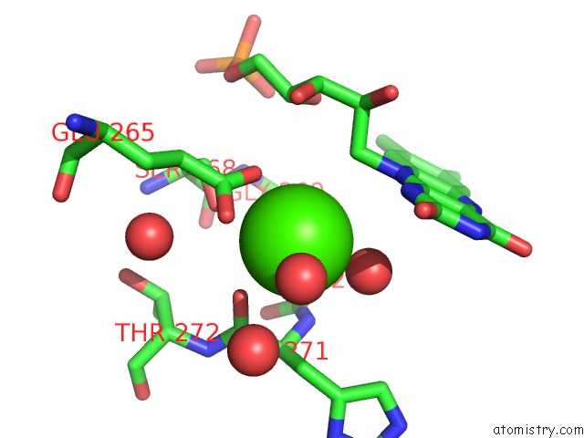
Mono view
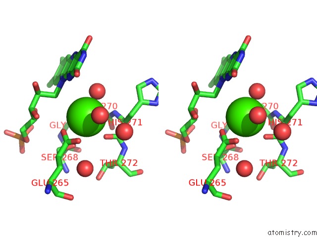
Stereo pair view
Mono view
Stereo pair view
A full contact list of Calcium with other atoms in the Ca binding
site number 2 of Crystal Structure of Pfad From Shewanella Piezotolerans in Complex with Fmn within 5.0Å range:
|
Reference:
M.L.Zhang,
H.D.Zhang,
Q.Li,
S.S.Meng,
L.J.Guo,
L.He,
J.Z.Huang,
L.Li.
Structural Insight Into the Enoyl Reduction in the Biosynthesis of Long-Chain Polyunsaturated Fatty Acid in Shewanella Piezotolerans To Be Published.
Page generated: Wed Jul 9 15:55:30 2025
Last articles
Mg in 7Y4AMg in 7Y44
Mg in 7Y43
Mg in 7XXF
Mg in 7Y3S
Mg in 7Y28
Mg in 7XYR
Mg in 7Y1W
Mg in 7Y1N
Mg in 7Y1M