Calcium »
PDB 6m5a-6mrm »
6mf2 »
Calcium in PDB 6mf2: Improved Model of Human Coagulation Factor VIII
Protein crystallography data
The structure of Improved Model of Human Coagulation Factor VIII, PDB code: 6mf2
was solved by
I.W.Smith,
P.C.Spiegel,
with X-Ray Crystallography technique. A brief refinement statistics is given in the table below:
| Resolution Low / High (Å) | 57.36 / 3.61 |
| Space group | P 41 21 2 |
| Cell size a, b, c (Å), α, β, γ (°) | 134.569, 134.569, 359.496, 90.00, 90.00, 90.00 |
| R / Rfree (%) | 25.2 / 28.4 |
Other elements in 6mf2:
The structure of Improved Model of Human Coagulation Factor VIII also contains other interesting chemical elements:
| Copper | (Cu) | 1 atom |
| Zinc | (Zn) | 1 atom |
Calcium Binding Sites:
The binding sites of Calcium atom in the Improved Model of Human Coagulation Factor VIII
(pdb code 6mf2). This binding sites where shown within
5.0 Angstroms radius around Calcium atom.
In total only one binding site of Calcium was determined in the Improved Model of Human Coagulation Factor VIII, PDB code: 6mf2:
In total only one binding site of Calcium was determined in the Improved Model of Human Coagulation Factor VIII, PDB code: 6mf2:
Calcium binding site 1 out of 1 in 6mf2
Go back to
Calcium binding site 1 out
of 1 in the Improved Model of Human Coagulation Factor VIII
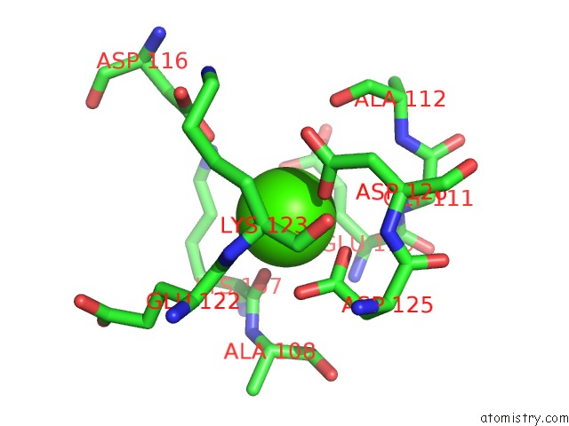
Mono view
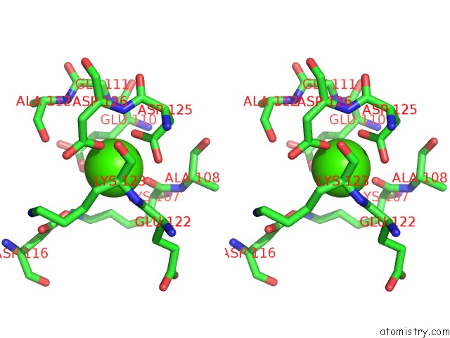
Stereo pair view
Mono view
Stereo pair view
A full contact list of Calcium with other atoms in the Ca binding
site number 1 of Improved Model of Human Coagulation Factor VIII within 5.0Å range:
|
Reference:
I.W.Smith,
A.E.D'aquino,
C.W.Coyle,
A.Fedanov,
E.T.Parker,
G.Denning,
H.T.Spencer,
P.Lollar,
C.B.Doering,
P.C.Spiegel Jr..
The 3.2 Angstrom Structure of A Bioengineered Variant of Blood Coagulation Factor VIII Indicates Two Conformations of the C2 Domain. J.Thromb.Haemost. 2019.
ISSN: ESSN 1538-7836
PubMed: 31454152
DOI: 10.1111/JTH.14621
Page generated: Wed Jul 9 16:06:08 2025
ISSN: ESSN 1538-7836
PubMed: 31454152
DOI: 10.1111/JTH.14621
Last articles
Zn in 5C0LZn in 5C0K
Zn in 5BZ0
Zn in 5BWZ
Zn in 5BYI
Zn in 5BWK
Zn in 5BWO
Zn in 5BWN
Zn in 5BWL
Zn in 5BUO