Calcium »
PDB 6mro-6n9d »
6n1a »
Calcium in PDB 6n1a: Crystal Structure of An N-Acetylgalactosamine Deacetylase From F. Plautii
Protein crystallography data
The structure of Crystal Structure of An N-Acetylgalactosamine Deacetylase From F. Plautii, PDB code: 6n1a
was solved by
L.Sim,
P.Rahfeld,
S.G.Withers,
with X-Ray Crystallography technique. A brief refinement statistics is given in the table below:
| Resolution Low / High (Å) | 46.20 / 1.60 |
| Space group | P 21 21 21 |
| Cell size a, b, c (Å), α, β, γ (°) | 51.584, 69.186, 104.325, 90.00, 90.00, 90.00 |
| R / Rfree (%) | 13.9 / 16.7 |
Other elements in 6n1a:
The structure of Crystal Structure of An N-Acetylgalactosamine Deacetylase From F. Plautii also contains other interesting chemical elements:
| Manganese | (Mn) | 1 atom |
Calcium Binding Sites:
The binding sites of Calcium atom in the Crystal Structure of An N-Acetylgalactosamine Deacetylase From F. Plautii
(pdb code 6n1a). This binding sites where shown within
5.0 Angstroms radius around Calcium atom.
In total 2 binding sites of Calcium where determined in the Crystal Structure of An N-Acetylgalactosamine Deacetylase From F. Plautii, PDB code: 6n1a:
Jump to Calcium binding site number: 1; 2;
In total 2 binding sites of Calcium where determined in the Crystal Structure of An N-Acetylgalactosamine Deacetylase From F. Plautii, PDB code: 6n1a:
Jump to Calcium binding site number: 1; 2;
Calcium binding site 1 out of 2 in 6n1a
Go back to
Calcium binding site 1 out
of 2 in the Crystal Structure of An N-Acetylgalactosamine Deacetylase From F. Plautii
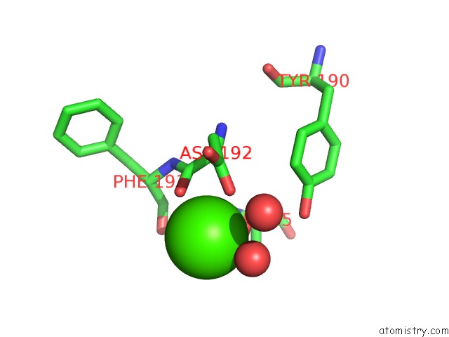
Mono view
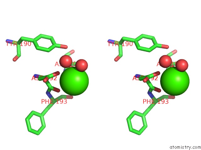
Stereo pair view
Mono view
Stereo pair view
A full contact list of Calcium with other atoms in the Ca binding
site number 1 of Crystal Structure of An N-Acetylgalactosamine Deacetylase From F. Plautii within 5.0Å range:
|
Calcium binding site 2 out of 2 in 6n1a
Go back to
Calcium binding site 2 out
of 2 in the Crystal Structure of An N-Acetylgalactosamine Deacetylase From F. Plautii
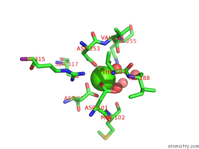
Mono view
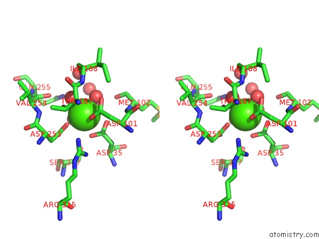
Stereo pair view
Mono view
Stereo pair view
A full contact list of Calcium with other atoms in the Ca binding
site number 2 of Crystal Structure of An N-Acetylgalactosamine Deacetylase From F. Plautii within 5.0Å range:
|
Reference:
P.Rahfeld,
L.Sim,
H.Moon,
I.Constantinescu,
C.Morgan-Lang,
S.J.Hallam,
J.N.Kizhakkedathu,
S.G.Withers.
An Enzymatic Pathway in the Human Gut Microbiome That Converts A to Universal O Type Blood. Nat Microbiol V. 4 1475 2019.
ISSN: ESSN 2058-5276
PubMed: 31182795
DOI: 10.1038/S41564-019-0469-7
Page generated: Wed Jul 9 16:12:52 2025
ISSN: ESSN 2058-5276
PubMed: 31182795
DOI: 10.1038/S41564-019-0469-7
Last articles
I in 3WN5I in 3WYX
I in 3WGW
I in 3WD6
I in 3WB5
I in 3W31
I in 3WB4
I in 3W1N
I in 3W0F
I in 3W2Z