Calcium »
PDB 6mro-6n9d »
6n22 »
Calcium in PDB 6n22: Crystal Structure of Mouse Protocadherin-15 EC1-2 Bap
Protein crystallography data
The structure of Crystal Structure of Mouse Protocadherin-15 EC1-2 Bap, PDB code: 6n22
was solved by
Y.Narui,
M.Sotomayor,
with X-Ray Crystallography technique. A brief refinement statistics is given in the table below:
| Resolution Low / High (Å) | 49.86 / 2.40 |
| Space group | P 64 |
| Cell size a, b, c (Å), α, β, γ (°) | 99.616, 99.616, 58.560, 90.00, 90.00, 120.00 |
| R / Rfree (%) | 17 / 23.3 |
Calcium Binding Sites:
The binding sites of Calcium atom in the Crystal Structure of Mouse Protocadherin-15 EC1-2 Bap
(pdb code 6n22). This binding sites where shown within
5.0 Angstroms radius around Calcium atom.
In total 3 binding sites of Calcium where determined in the Crystal Structure of Mouse Protocadherin-15 EC1-2 Bap, PDB code: 6n22:
Jump to Calcium binding site number: 1; 2; 3;
In total 3 binding sites of Calcium where determined in the Crystal Structure of Mouse Protocadherin-15 EC1-2 Bap, PDB code: 6n22:
Jump to Calcium binding site number: 1; 2; 3;
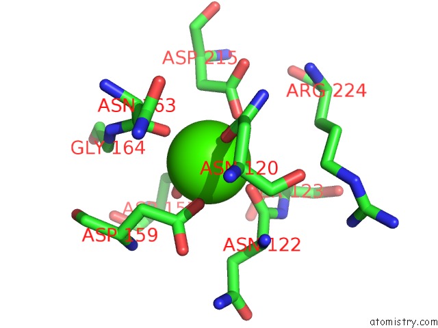
Calcium binding site 1 out of 3 in 6n22
Go back to
Calcium binding site 1 out
of 3 in the Crystal Structure of Mouse Protocadherin-15 EC1-2 Bap
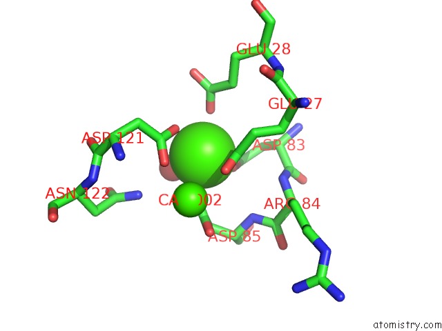
Mono view
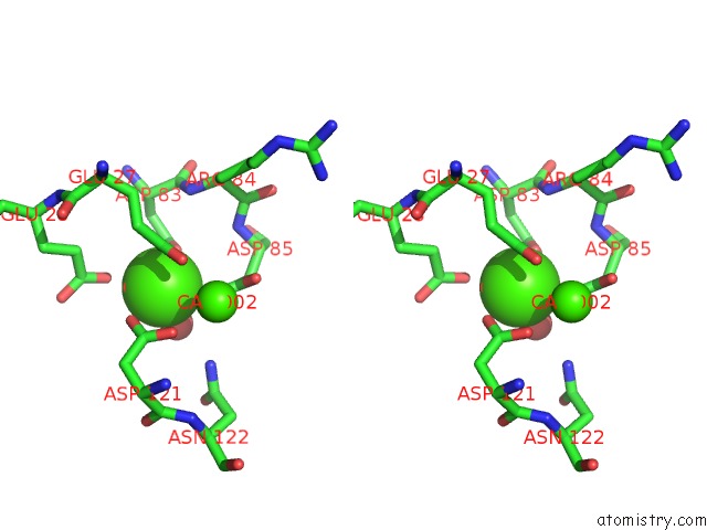
Stereo pair view
Mono view
Stereo pair view
A full contact list of Calcium with other atoms in the Ca binding
site number 1 of Crystal Structure of Mouse Protocadherin-15 EC1-2 Bap within 5.0Å range:
|
Calcium binding site 2 out of 3 in 6n22
Go back to
Calcium binding site 2 out
of 3 in the Crystal Structure of Mouse Protocadherin-15 EC1-2 Bap
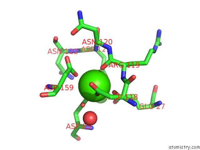
Mono view
Stereo pair view
Mono view
Stereo pair view
A full contact list of Calcium with other atoms in the Ca binding
site number 2 of Crystal Structure of Mouse Protocadherin-15 EC1-2 Bap within 5.0Å range:
|
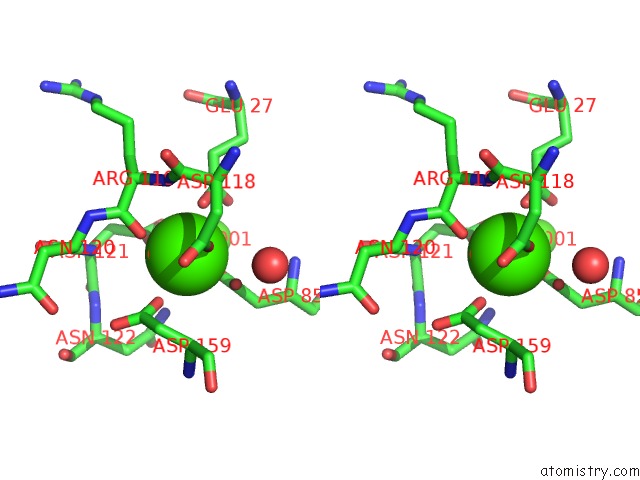
Calcium binding site 3 out of 3 in 6n22
Go back to
Calcium binding site 3 out
of 3 in the Crystal Structure of Mouse Protocadherin-15 EC1-2 Bap
Mono view

Stereo pair view
Mono view
Stereo pair view
A full contact list of Calcium with other atoms in the Ca binding
site number 3 of Crystal Structure of Mouse Protocadherin-15 EC1-2 Bap within 5.0Å range:
|
Reference:
Y.Narui,
M.Sotomayor.
Crystal Structure of Mouse Protocadherin-15 EC1-2 Bap To Be Published.
Page generated: Wed Jul 9 16:13:01 2025
Last articles
I in 3WN5I in 3WYX
I in 3WGW
I in 3WD6
I in 3WB5
I in 3W31
I in 3WB4
I in 3W1N
I in 3W0F
I in 3W2Z