Calcium »
PDB 6n9m-6o20 »
6nvx »
Calcium in PDB 6nvx: Crystal Structure of Penicillin G Acylase From Bacillus Sp. Fjat-27231
Protein crystallography data
The structure of Crystal Structure of Penicillin G Acylase From Bacillus Sp. Fjat-27231, PDB code: 6nvx
was solved by
W.Blankenfeldt,
with X-Ray Crystallography technique. A brief refinement statistics is given in the table below:
| Resolution Low / High (Å) | 46.15 / 1.36 |
| Space group | P 21 21 21 |
| Cell size a, b, c (Å), α, β, γ (°) | 57.366, 77.686, 210.365, 90.00, 90.00, 90.00 |
| R / Rfree (%) | 14.9 / 16.7 |
Calcium Binding Sites:
The binding sites of Calcium atom in the Crystal Structure of Penicillin G Acylase From Bacillus Sp. Fjat-27231
(pdb code 6nvx). This binding sites where shown within
5.0 Angstroms radius around Calcium atom.
In total 2 binding sites of Calcium where determined in the Crystal Structure of Penicillin G Acylase From Bacillus Sp. Fjat-27231, PDB code: 6nvx:
Jump to Calcium binding site number: 1; 2;
In total 2 binding sites of Calcium where determined in the Crystal Structure of Penicillin G Acylase From Bacillus Sp. Fjat-27231, PDB code: 6nvx:
Jump to Calcium binding site number: 1; 2;
Calcium binding site 1 out of 2 in 6nvx
Go back to
Calcium binding site 1 out
of 2 in the Crystal Structure of Penicillin G Acylase From Bacillus Sp. Fjat-27231
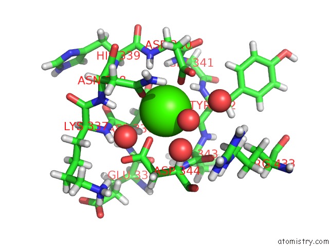
Mono view
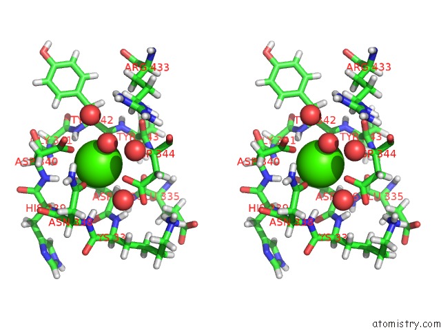
Stereo pair view
Mono view
Stereo pair view
A full contact list of Calcium with other atoms in the Ca binding
site number 1 of Crystal Structure of Penicillin G Acylase From Bacillus Sp. Fjat-27231 within 5.0Å range:
|
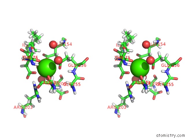
Calcium binding site 2 out of 2 in 6nvx
Go back to
Calcium binding site 2 out
of 2 in the Crystal Structure of Penicillin G Acylase From Bacillus Sp. Fjat-27231
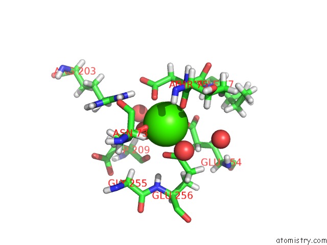
Mono view
Stereo pair view
Mono view
Stereo pair view
A full contact list of Calcium with other atoms in the Ca binding
site number 2 of Crystal Structure of Penicillin G Acylase From Bacillus Sp. Fjat-27231 within 5.0Å range:
|
Reference:
J.Mayer,
J.Pippel,
G.Gunther,
C.Muller,
A.Lauermann,
T.Knuuti,
W.Blankenfeldt,
D.Jahn,
R.Biedendieck.
Crystal Structures and Protein Engineering of Three Different Penicillin G Acylases From Gram-Positive Bacteria with Different Thermostability. Appl.Microbiol.Biotechnol. V. 103 7537 2019.
ISSN: ESSN 1432-0614
PubMed: 31227867
DOI: 10.1007/S00253-019-09977-8
Page generated: Wed Jul 9 16:23:27 2025
ISSN: ESSN 1432-0614
PubMed: 31227867
DOI: 10.1007/S00253-019-09977-8
Last articles
Mg in 5XF5Mg in 5XF4
Mg in 5XF3
Mg in 5XDX
Mg in 5XD5
Mg in 5XE5
Mg in 5XDQ
Mg in 5XDR
Mg in 5XD9
Mg in 5XD8