Calcium »
PDB 6o44-6ok8 »
6o44 »
Calcium in PDB 6o44: Insight Into Subtilisin E-S7 Cleavage Pattern Based on Crystal Structure and Hydrolysates Peptide Analysis
Enzymatic activity of Insight Into Subtilisin E-S7 Cleavage Pattern Based on Crystal Structure and Hydrolysates Peptide Analysis
All present enzymatic activity of Insight Into Subtilisin E-S7 Cleavage Pattern Based on Crystal Structure and Hydrolysates Peptide Analysis:
3.4.21.62;
3.4.21.62;
Protein crystallography data
The structure of Insight Into Subtilisin E-S7 Cleavage Pattern Based on Crystal Structure and Hydrolysates Peptide Analysis, PDB code: 6o44
was solved by
H.Tang,
K.Shi,
H.Aihara,
with X-Ray Crystallography technique. A brief refinement statistics is given in the table below:
| Resolution Low / High (Å) | 59.16 / 1.83 |
| Space group | P 21 21 21 |
| Cell size a, b, c (Å), α, β, γ (°) | 74.177, 80.123, 87.729, 90.00, 90.00, 90.00 |
| R / Rfree (%) | 18.7 / 22.2 |
Calcium Binding Sites:
The binding sites of Calcium atom in the Insight Into Subtilisin E-S7 Cleavage Pattern Based on Crystal Structure and Hydrolysates Peptide Analysis
(pdb code 6o44). This binding sites where shown within
5.0 Angstroms radius around Calcium atom.
In total 4 binding sites of Calcium where determined in the Insight Into Subtilisin E-S7 Cleavage Pattern Based on Crystal Structure and Hydrolysates Peptide Analysis, PDB code: 6o44:
Jump to Calcium binding site number: 1; 2; 3; 4;
In total 4 binding sites of Calcium where determined in the Insight Into Subtilisin E-S7 Cleavage Pattern Based on Crystal Structure and Hydrolysates Peptide Analysis, PDB code: 6o44:
Jump to Calcium binding site number: 1; 2; 3; 4;
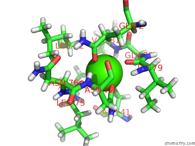
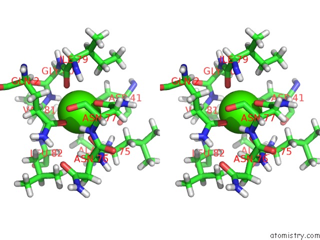
Calcium binding site 1 out of 4 in 6o44
Go back to
Calcium binding site 1 out
of 4 in the Insight Into Subtilisin E-S7 Cleavage Pattern Based on Crystal Structure and Hydrolysates Peptide Analysis
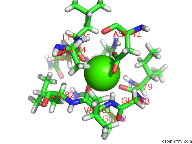
Mono view
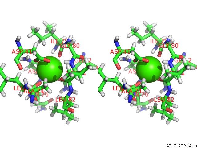
Stereo pair view
Mono view
Stereo pair view
A full contact list of Calcium with other atoms in the Ca binding
site number 1 of Insight Into Subtilisin E-S7 Cleavage Pattern Based on Crystal Structure and Hydrolysates Peptide Analysis within 5.0Å range:
|
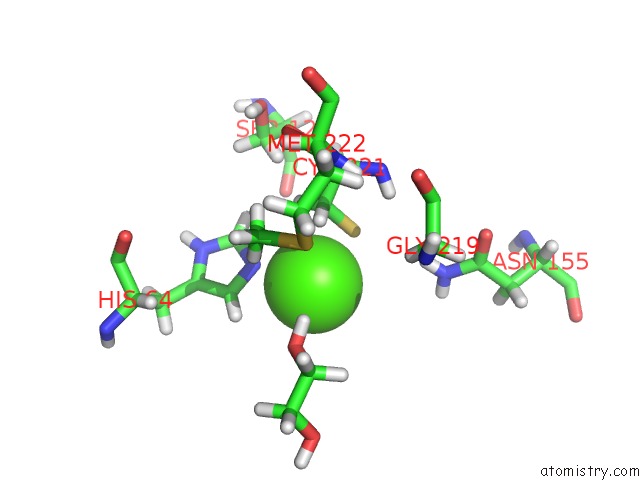
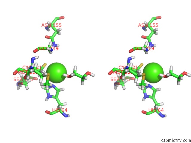
Calcium binding site 2 out of 4 in 6o44
Go back to
Calcium binding site 2 out
of 4 in the Insight Into Subtilisin E-S7 Cleavage Pattern Based on Crystal Structure and Hydrolysates Peptide Analysis
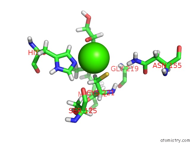
Mono view
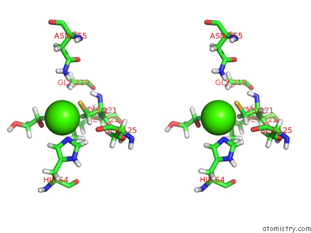
Stereo pair view
Mono view
Stereo pair view
A full contact list of Calcium with other atoms in the Ca binding
site number 2 of Insight Into Subtilisin E-S7 Cleavage Pattern Based on Crystal Structure and Hydrolysates Peptide Analysis within 5.0Å range:
|
Calcium binding site 3 out of 4 in 6o44
Go back to
Calcium binding site 3 out
of 4 in the Insight Into Subtilisin E-S7 Cleavage Pattern Based on Crystal Structure and Hydrolysates Peptide Analysis
Mono view
Stereo pair view
Mono view
Stereo pair view
A full contact list of Calcium with other atoms in the Ca binding
site number 3 of Insight Into Subtilisin E-S7 Cleavage Pattern Based on Crystal Structure and Hydrolysates Peptide Analysis within 5.0Å range:
|
Calcium binding site 4 out of 4 in 6o44
Go back to
Calcium binding site 4 out
of 4 in the Insight Into Subtilisin E-S7 Cleavage Pattern Based on Crystal Structure and Hydrolysates Peptide Analysis
Mono view
Stereo pair view
Mono view
Stereo pair view
A full contact list of Calcium with other atoms in the Ca binding
site number 4 of Insight Into Subtilisin E-S7 Cleavage Pattern Based on Crystal Structure and Hydrolysates Peptide Analysis within 5.0Å range:
|
Reference:
H.Tang,
J.Zhang,
K.Shi,
H.Aihara,
G.Du.
Insight Into Subtilisin E-S7 Cleavage Pattern Based on Crystal Structure and Hydrolysates Peptide Analysis. Biochem. Biophys. Res. V. 512 623 2019COMMUN..
ISSN: ESSN 1090-2104
PubMed: 30914195
DOI: 10.1016/J.BBRC.2019.03.064
Page generated: Wed Jul 9 16:25:20 2025
ISSN: ESSN 1090-2104
PubMed: 30914195
DOI: 10.1016/J.BBRC.2019.03.064
Last articles
Mg in 6TRAMg in 6TR4
Mg in 6TR3
Mg in 6TMF
Mg in 6TQO
Mg in 6TQN
Mg in 6TQF
Mg in 6TQE
Mg in 6TQB
Mg in 6TQA