Calcium »
PDB 6o44-6ok8 »
6ohh »
Calcium in PDB 6ohh: Structure of EF1P2_MFAP2B Bound to Dfhbi
Protein crystallography data
The structure of Structure of EF1P2_MFAP2B Bound to Dfhbi, PDB code: 6ohh
was solved by
L.A.Doyle,
B.L.Stoddard,
with X-Ray Crystallography technique. A brief refinement statistics is given in the table below:
| Resolution Low / High (Å) | 25.42 / 2.10 |
| Space group | P 1 21 1 |
| Cell size a, b, c (Å), α, β, γ (°) | 36.350, 35.564, 86.596, 90.00, 90.68, 90.00 |
| R / Rfree (%) | 16.9 / 20.1 |
Other elements in 6ohh:
The structure of Structure of EF1P2_MFAP2B Bound to Dfhbi also contains other interesting chemical elements:
| Fluorine | (F) | 4 atoms |
Calcium Binding Sites:
The binding sites of Calcium atom in the Structure of EF1P2_MFAP2B Bound to Dfhbi
(pdb code 6ohh). This binding sites where shown within
5.0 Angstroms radius around Calcium atom.
In total 2 binding sites of Calcium where determined in the Structure of EF1P2_MFAP2B Bound to Dfhbi, PDB code: 6ohh:
Jump to Calcium binding site number: 1; 2;
In total 2 binding sites of Calcium where determined in the Structure of EF1P2_MFAP2B Bound to Dfhbi, PDB code: 6ohh:
Jump to Calcium binding site number: 1; 2;
Calcium binding site 1 out of 2 in 6ohh
Go back to
Calcium binding site 1 out
of 2 in the Structure of EF1P2_MFAP2B Bound to Dfhbi
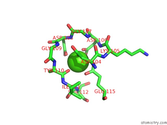
Mono view
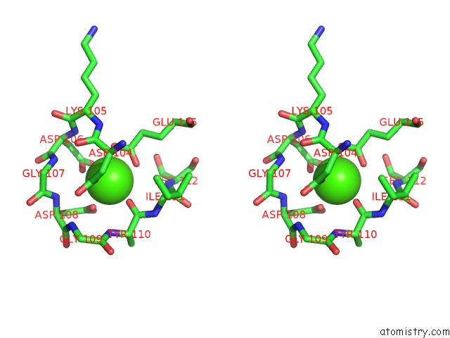
Stereo pair view
Mono view
Stereo pair view
A full contact list of Calcium with other atoms in the Ca binding
site number 1 of Structure of EF1P2_MFAP2B Bound to Dfhbi within 5.0Å range:
|
Calcium binding site 2 out of 2 in 6ohh
Go back to
Calcium binding site 2 out
of 2 in the Structure of EF1P2_MFAP2B Bound to Dfhbi
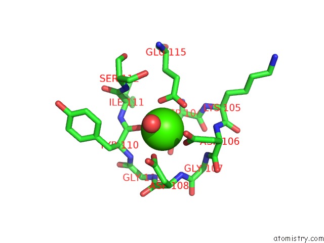
Mono view
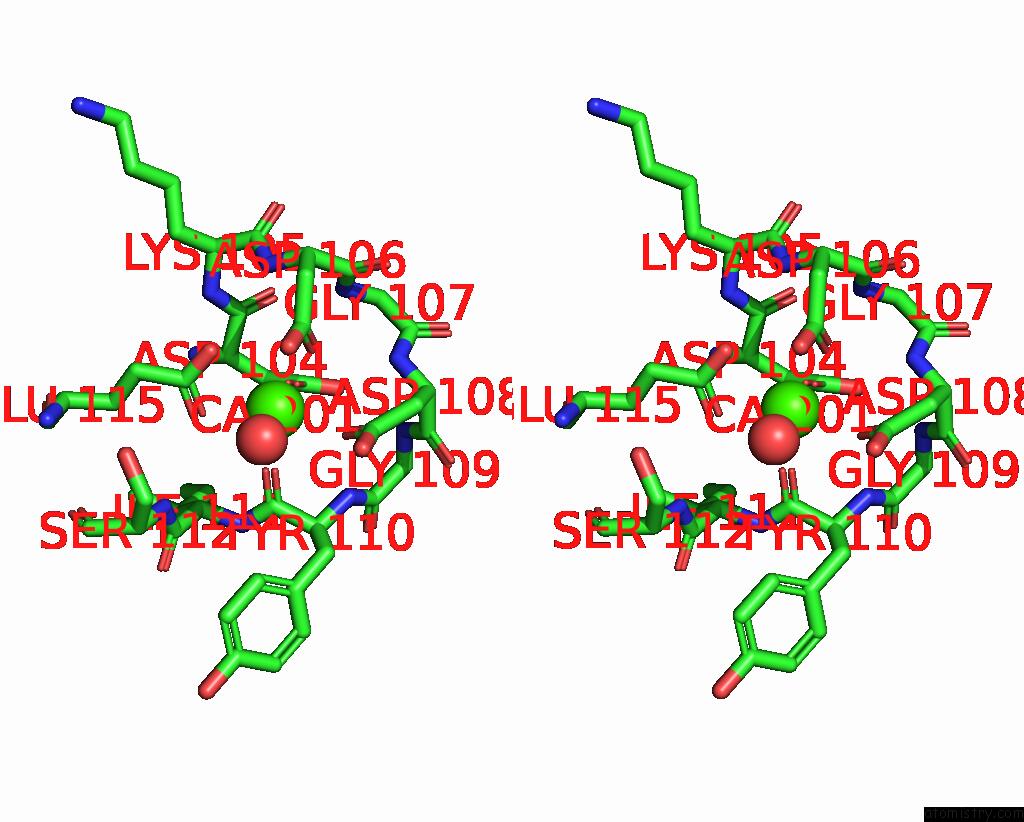
Stereo pair view
Mono view
Stereo pair view
A full contact list of Calcium with other atoms in the Ca binding
site number 2 of Structure of EF1P2_MFAP2B Bound to Dfhbi within 5.0Å range:
|
Reference:
L.A.Doyle,
L.A.Doyle,
B.L.Stoddard.
N/A N/A.
Page generated: Wed Jul 9 16:33:28 2025
Last articles
Mg in 6YKWMg in 6YKV
Mg in 6YKU
Mg in 6YKT
Mg in 6YKS
Mg in 6YKQ
Mg in 6YKO
Mg in 6YKN
Mg in 6YKL
Mg in 6YKK