Calcium »
PDB 6p8z-6pu5 »
6pmp »
Calcium in PDB 6pmp: Crystal Structure of A Fragment of Rat Phospholipase Cepsilon EF3-RA1
Enzymatic activity of Crystal Structure of A Fragment of Rat Phospholipase Cepsilon EF3-RA1
All present enzymatic activity of Crystal Structure of A Fragment of Rat Phospholipase Cepsilon EF3-RA1:
3.1.4.11;
3.1.4.11;
Protein crystallography data
The structure of Crystal Structure of A Fragment of Rat Phospholipase Cepsilon EF3-RA1, PDB code: 6pmp
was solved by
N.Y.Rugema,
A.M.Lyon,
with X-Ray Crystallography technique. A brief refinement statistics is given in the table below:
| Resolution Low / High (Å) | 20.00 / 2.73 |
| Space group | P 1 21 1 |
| Cell size a, b, c (Å), α, β, γ (°) | 93.572, 127.755, 139.337, 90.00, 101.12, 90.00 |
| R / Rfree (%) | 23.4 / 27.3 |
Calcium Binding Sites:
The binding sites of Calcium atom in the Crystal Structure of A Fragment of Rat Phospholipase Cepsilon EF3-RA1
(pdb code 6pmp). This binding sites where shown within
5.0 Angstroms radius around Calcium atom.
In total 4 binding sites of Calcium where determined in the Crystal Structure of A Fragment of Rat Phospholipase Cepsilon EF3-RA1, PDB code: 6pmp:
Jump to Calcium binding site number: 1; 2; 3; 4;
In total 4 binding sites of Calcium where determined in the Crystal Structure of A Fragment of Rat Phospholipase Cepsilon EF3-RA1, PDB code: 6pmp:
Jump to Calcium binding site number: 1; 2; 3; 4;
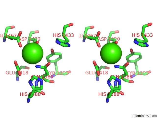
Calcium binding site 1 out of 4 in 6pmp
Go back to
Calcium binding site 1 out
of 4 in the Crystal Structure of A Fragment of Rat Phospholipase Cepsilon EF3-RA1
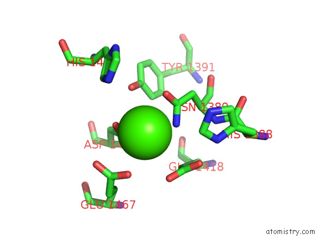
Mono view
Stereo pair view
Mono view
Stereo pair view
A full contact list of Calcium with other atoms in the Ca binding
site number 1 of Crystal Structure of A Fragment of Rat Phospholipase Cepsilon EF3-RA1 within 5.0Å range:
|
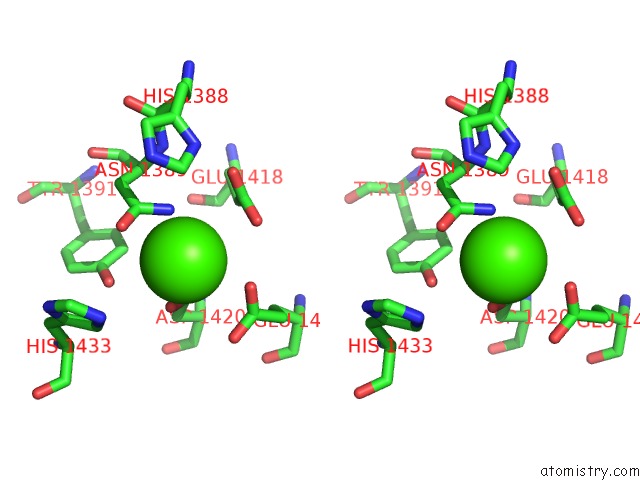
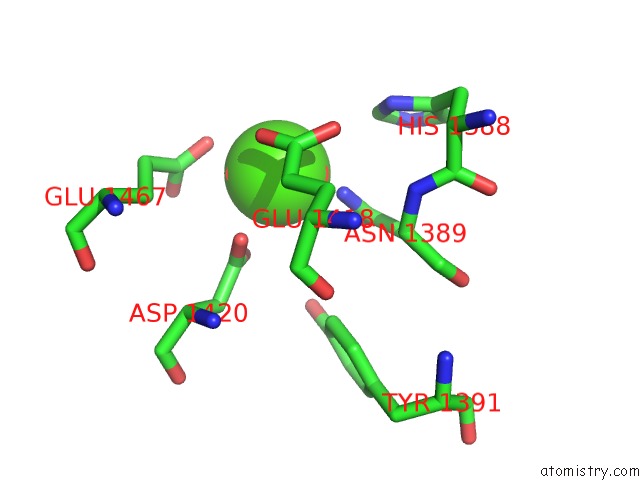
Calcium binding site 2 out of 4 in 6pmp
Go back to
Calcium binding site 2 out
of 4 in the Crystal Structure of A Fragment of Rat Phospholipase Cepsilon EF3-RA1

Mono view
Stereo pair view
Mono view
Stereo pair view
A full contact list of Calcium with other atoms in the Ca binding
site number 2 of Crystal Structure of A Fragment of Rat Phospholipase Cepsilon EF3-RA1 within 5.0Å range:
|
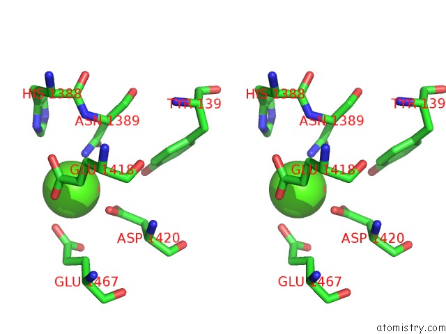
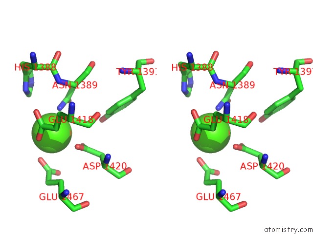
Calcium binding site 3 out of 4 in 6pmp
Go back to
Calcium binding site 3 out
of 4 in the Crystal Structure of A Fragment of Rat Phospholipase Cepsilon EF3-RA1
Mono view
Stereo pair view
Mono view
Stereo pair view
A full contact list of Calcium with other atoms in the Ca binding
site number 3 of Crystal Structure of A Fragment of Rat Phospholipase Cepsilon EF3-RA1 within 5.0Å range:
|
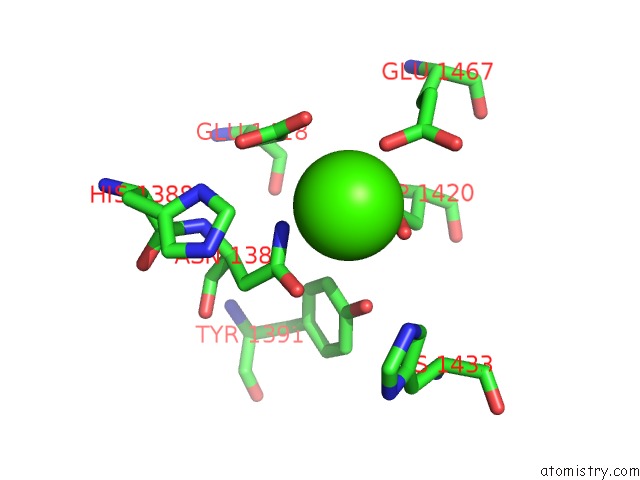
Calcium binding site 4 out of 4 in 6pmp
Go back to
Calcium binding site 4 out
of 4 in the Crystal Structure of A Fragment of Rat Phospholipase Cepsilon EF3-RA1
Mono view
Stereo pair view
Mono view
Stereo pair view
A full contact list of Calcium with other atoms in the Ca binding
site number 4 of Crystal Structure of A Fragment of Rat Phospholipase Cepsilon EF3-RA1 within 5.0Å range:
|
Reference:
N.Y.Rugema,
E.E.Garland-Kuntz,
M.Sieng,
K.Muralidharan,
M.M.Van Camp,
H.O'neill,
W.Mbongo,
A.F.Selvia,
A.T.Marti,
A.Everly,
E.Mckenzie,
A.M.Lyon.
Structure of Phospholipase C Epsilon Reveals An Integrated RA1 Domain and Previously Unidentified Regulatory Elements. Commun Biol V. 3 445 2020.
ISSN: ESSN 2399-3642
PubMed: 32796910
DOI: 10.1038/S42003-020-01178-8
Page generated: Wed Jul 9 16:48:48 2025
ISSN: ESSN 2399-3642
PubMed: 32796910
DOI: 10.1038/S42003-020-01178-8
Last articles
Mg in 2BWTMg in 2BVN
Mg in 2BV3
Mg in 2BUP
Mg in 2BTD
Mg in 2BU8
Mg in 2BU2
Mg in 2BT6
Mg in 2BHW
Mg in 2BT1