Calcium »
PDB 6pus-6q79 »
6puv »
Calcium in PDB 6puv: Structure of the Crd of CLEC10A
Protein crystallography data
The structure of Structure of the Crd of CLEC10A, PDB code: 6puv
was solved by
G.Birrane,
P.V.Murphy,
A.Gabba,
J.G.Luz,
with X-Ray Crystallography technique. A brief refinement statistics is given in the table below:
| Resolution Low / High (Å) | 45.00 / 1.20 |
| Space group | P 21 21 21 |
| Cell size a, b, c (Å), α, β, γ (°) | 28.958, 42.701, 89.958, 90.00, 90.00, 90.00 |
| R / Rfree (%) | 15.5 / 18.9 |
Calcium Binding Sites:
The binding sites of Calcium atom in the Structure of the Crd of CLEC10A
(pdb code 6puv). This binding sites where shown within
5.0 Angstroms radius around Calcium atom.
In total only one binding site of Calcium was determined in the Structure of the Crd of CLEC10A, PDB code: 6puv:
In total only one binding site of Calcium was determined in the Structure of the Crd of CLEC10A, PDB code: 6puv:
Calcium binding site 1 out of 1 in 6puv
Go back to
Calcium binding site 1 out
of 1 in the Structure of the Crd of CLEC10A
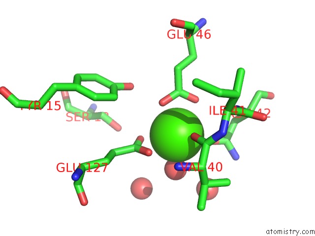
Mono view
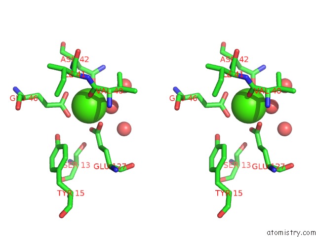
Stereo pair view
Mono view
Stereo pair view
A full contact list of Calcium with other atoms in the Ca binding
site number 1 of Structure of the Crd of CLEC10A within 5.0Å range:
|
Reference:
G.Birrane,
P.V.Murphy.
Structure of the Carbohydrate Recognition Domain of CLEC10A To Be Published.
Page generated: Wed Jul 9 16:55:09 2025
Last articles
Mg in 4DV3Mg in 4DV2
Mg in 4DV0
Mg in 4DV1
Mg in 4DUZ
Mg in 4DUY
Mg in 4DR7
Mg in 4DR6
Mg in 4DR5
Mg in 4DUX