Calcium »
PDB 6q7o-6qps »
6q86 »
Calcium in PDB 6q86: Structure of Fucosylated D-Antimicrobial Peptide SB4 in Complex with the Fucose-Binding Lectin Pa-Iil at 2.008 Angstrom Resolution
Protein crystallography data
The structure of Structure of Fucosylated D-Antimicrobial Peptide SB4 in Complex with the Fucose-Binding Lectin Pa-Iil at 2.008 Angstrom Resolution, PDB code: 6q86
was solved by
S.Baeriswyl,
A.Stocker,
J.L.Reymond,
with X-Ray Crystallography technique. A brief refinement statistics is given in the table below:
| Resolution Low / High (Å) | 48.15 / 2.01 |
| Space group | P 31 2 1 |
| Cell size a, b, c (Å), α, β, γ (°) | 55.600, 55.600, 150.621, 90.00, 90.00, 120.00 |
| R / Rfree (%) | 15.7 / 19 |
Calcium Binding Sites:
The binding sites of Calcium atom in the Structure of Fucosylated D-Antimicrobial Peptide SB4 in Complex with the Fucose-Binding Lectin Pa-Iil at 2.008 Angstrom Resolution
(pdb code 6q86). This binding sites where shown within
5.0 Angstroms radius around Calcium atom.
In total 4 binding sites of Calcium where determined in the Structure of Fucosylated D-Antimicrobial Peptide SB4 in Complex with the Fucose-Binding Lectin Pa-Iil at 2.008 Angstrom Resolution, PDB code: 6q86:
Jump to Calcium binding site number: 1; 2; 3; 4;
In total 4 binding sites of Calcium where determined in the Structure of Fucosylated D-Antimicrobial Peptide SB4 in Complex with the Fucose-Binding Lectin Pa-Iil at 2.008 Angstrom Resolution, PDB code: 6q86:
Jump to Calcium binding site number: 1; 2; 3; 4;
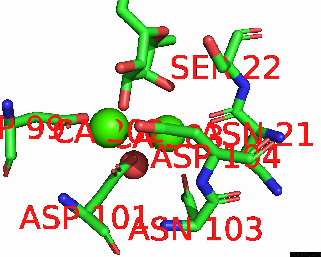
Calcium binding site 1 out of 4 in 6q86
Go back to
Calcium binding site 1 out
of 4 in the Structure of Fucosylated D-Antimicrobial Peptide SB4 in Complex with the Fucose-Binding Lectin Pa-Iil at 2.008 Angstrom Resolution
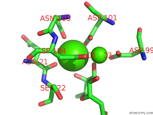
Mono view
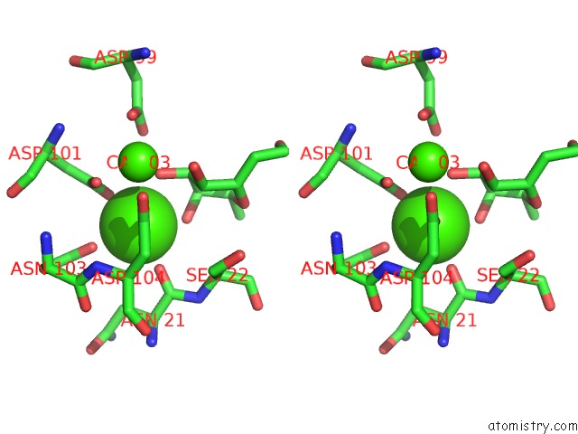
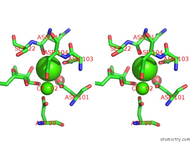
Stereo pair view
Mono view
Stereo pair view
A full contact list of Calcium with other atoms in the Ca binding
site number 1 of Structure of Fucosylated D-Antimicrobial Peptide SB4 in Complex with the Fucose-Binding Lectin Pa-Iil at 2.008 Angstrom Resolution within 5.0Å range:
|
Calcium binding site 2 out of 4 in 6q86
Go back to
Calcium binding site 2 out
of 4 in the Structure of Fucosylated D-Antimicrobial Peptide SB4 in Complex with the Fucose-Binding Lectin Pa-Iil at 2.008 Angstrom Resolution

Mono view
Stereo pair view
Mono view
Stereo pair view
A full contact list of Calcium with other atoms in the Ca binding
site number 2 of Structure of Fucosylated D-Antimicrobial Peptide SB4 in Complex with the Fucose-Binding Lectin Pa-Iil at 2.008 Angstrom Resolution within 5.0Å range:
|
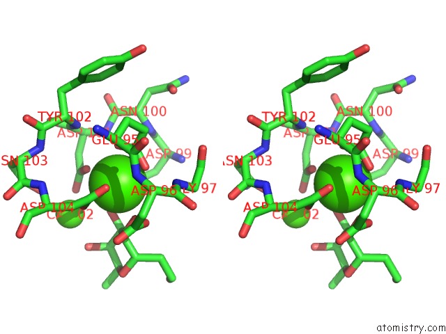
Calcium binding site 3 out of 4 in 6q86
Go back to
Calcium binding site 3 out
of 4 in the Structure of Fucosylated D-Antimicrobial Peptide SB4 in Complex with the Fucose-Binding Lectin Pa-Iil at 2.008 Angstrom Resolution
Mono view

Stereo pair view
Mono view
Stereo pair view
A full contact list of Calcium with other atoms in the Ca binding
site number 3 of Structure of Fucosylated D-Antimicrobial Peptide SB4 in Complex with the Fucose-Binding Lectin Pa-Iil at 2.008 Angstrom Resolution within 5.0Å range:
|
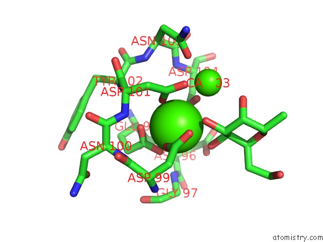
Calcium binding site 4 out of 4 in 6q86
Go back to
Calcium binding site 4 out
of 4 in the Structure of Fucosylated D-Antimicrobial Peptide SB4 in Complex with the Fucose-Binding Lectin Pa-Iil at 2.008 Angstrom Resolution
Mono view
Stereo pair view
Mono view
Stereo pair view
A full contact list of Calcium with other atoms in the Ca binding
site number 4 of Structure of Fucosylated D-Antimicrobial Peptide SB4 in Complex with the Fucose-Binding Lectin Pa-Iil at 2.008 Angstrom Resolution within 5.0Å range:
|
Reference:
S.Baeriswyl,
B.H.Gan,
T.N.Siriwardena,
R.Visini,
M.Robadey,
S.Javor,
A.Stocker,
T.Darbre,
J.L.Reymond.
X-Ray Crystal Structures of Short Antimicrobial Peptides As Pseudomonas Aeruginosa Lectin B Complexes. Acs Chem.Biol. V. 14 758 2019.
ISSN: ESSN 1554-8937
PubMed: 30830745
DOI: 10.1021/ACSCHEMBIO.9B00047
Page generated: Wed Jul 9 17:06:58 2025
ISSN: ESSN 1554-8937
PubMed: 30830745
DOI: 10.1021/ACSCHEMBIO.9B00047
Last articles
K in 2FRZK in 2FPQ
K in 2FPM
K in 2FPL
K in 2FPK
K in 2FPG
K in 2FP4
K in 2FEU
K in 2FFY
K in 2FDE