Calcium »
PDB 6q7o-6qps »
6qdt »
Calcium in PDB 6qdt: Crystal Structure of 14-3-3SIGMA in Complex with A RAPGEF2 PT740 Phosphopeptide
Protein crystallography data
The structure of Crystal Structure of 14-3-3SIGMA in Complex with A RAPGEF2 PT740 Phosphopeptide, PDB code: 6qdt
was solved by
S.A.Andrei,
A.Kaplan,
A.E.Fournier,
C.Ottman,
with X-Ray Crystallography technique. A brief refinement statistics is given in the table below:
| Resolution Low / High (Å) | 41.87 / 1.70 |
| Space group | C 2 2 21 |
| Cell size a, b, c (Å), α, β, γ (°) | 82.657, 112.411, 62.749, 90.00, 90.00, 90.00 |
| R / Rfree (%) | 16.2 / 19.2 |
Other elements in 6qdt:
The structure of Crystal Structure of 14-3-3SIGMA in Complex with A RAPGEF2 PT740 Phosphopeptide also contains other interesting chemical elements:
| Chlorine | (Cl) | 1 atom |
| Sodium | (Na) | 5 atoms |
Calcium Binding Sites:
The binding sites of Calcium atom in the Crystal Structure of 14-3-3SIGMA in Complex with A RAPGEF2 PT740 Phosphopeptide
(pdb code 6qdt). This binding sites where shown within
5.0 Angstroms radius around Calcium atom.
In total only one binding site of Calcium was determined in the Crystal Structure of 14-3-3SIGMA in Complex with A RAPGEF2 PT740 Phosphopeptide, PDB code: 6qdt:
In total only one binding site of Calcium was determined in the Crystal Structure of 14-3-3SIGMA in Complex with A RAPGEF2 PT740 Phosphopeptide, PDB code: 6qdt:
Calcium binding site 1 out of 1 in 6qdt
Go back to
Calcium binding site 1 out
of 1 in the Crystal Structure of 14-3-3SIGMA in Complex with A RAPGEF2 PT740 Phosphopeptide
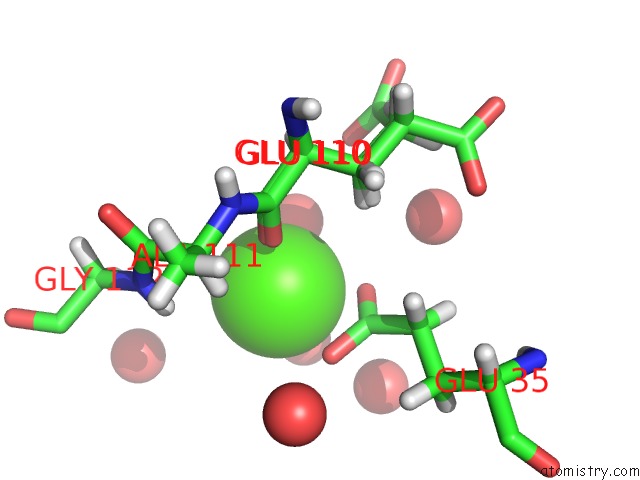
Mono view
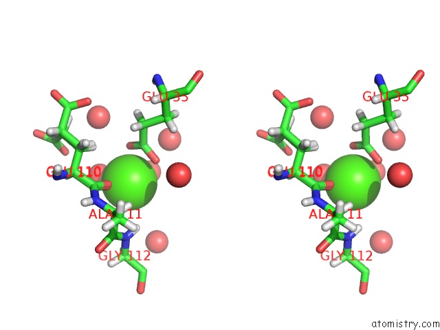
Stereo pair view
Mono view
Stereo pair view
A full contact list of Calcium with other atoms in the Ca binding
site number 1 of Crystal Structure of 14-3-3SIGMA in Complex with A RAPGEF2 PT740 Phosphopeptide within 5.0Å range:
|
Reference:
A.Kaplan,
S.A.Andrei,
C.Ottmann,
A.E.Fournier.
A Single-Agent Polypharmacological Approach to Stimulate Axon Regeneration To Be Published.
Page generated: Wed Jul 9 17:09:49 2025
Last articles
K in 7AQ5K in 7AQ4
K in 7AQ3
K in 7AOA
K in 7AQ2
K in 7AQ0
K in 7APY
K in 7AL9
K in 7AO9
K in 7AO8