Calcium »
PDB 6qq5-6r7u »
6r2q »
Calcium in PDB 6r2q: Structure of the Mtr Complex
Protein crystallography data
The structure of Structure of the Mtr Complex, PDB code: 6r2q
was solved by
T.A.Clarke,
M.J.Edwards,
with X-Ray Crystallography technique. A brief refinement statistics is given in the table below:
| Resolution Low / High (Å) | 73.25 / 2.70 |
| Space group | C 2 2 21 |
| Cell size a, b, c (Å), α, β, γ (°) | 212.042, 234.165, 99.192, 90.00, 90.00, 90.00 |
| R / Rfree (%) | 22.4 / 25.7 |
Other elements in 6r2q:
The structure of Structure of the Mtr Complex also contains other interesting chemical elements:
| Iron | (Fe) | 20 atoms |
Calcium Binding Sites:
The binding sites of Calcium atom in the Structure of the Mtr Complex
(pdb code 6r2q). This binding sites where shown within
5.0 Angstroms radius around Calcium atom.
In total 4 binding sites of Calcium where determined in the Structure of the Mtr Complex, PDB code: 6r2q:
Jump to Calcium binding site number: 1; 2; 3; 4;
In total 4 binding sites of Calcium where determined in the Structure of the Mtr Complex, PDB code: 6r2q:
Jump to Calcium binding site number: 1; 2; 3; 4;
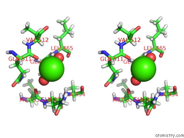
Calcium binding site 1 out of 4 in 6r2q
Go back to
Calcium binding site 1 out
of 4 in the Structure of the Mtr Complex
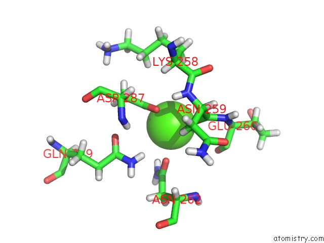
Mono view
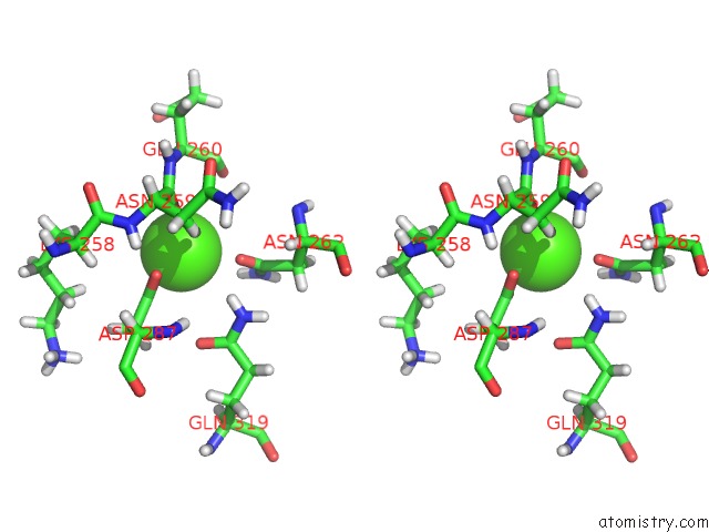
Stereo pair view
Mono view
Stereo pair view
A full contact list of Calcium with other atoms in the Ca binding
site number 1 of Structure of the Mtr Complex within 5.0Å range:
|
Calcium binding site 2 out of 4 in 6r2q
Go back to
Calcium binding site 2 out
of 4 in the Structure of the Mtr Complex

Mono view
Stereo pair view
Mono view
Stereo pair view
A full contact list of Calcium with other atoms in the Ca binding
site number 2 of Structure of the Mtr Complex within 5.0Å range:
|
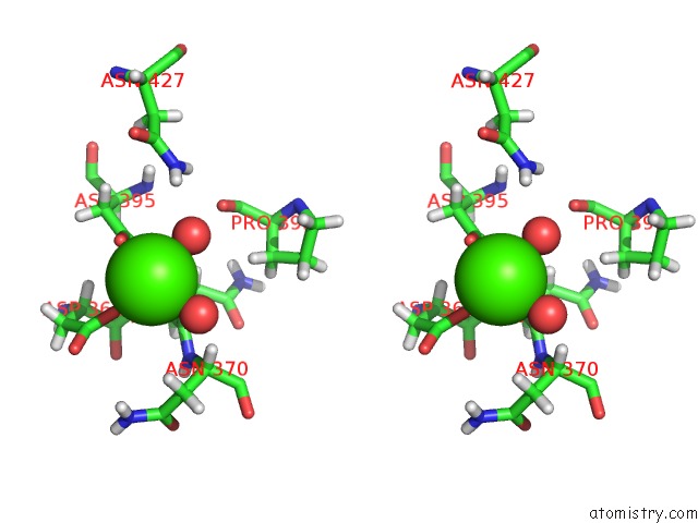
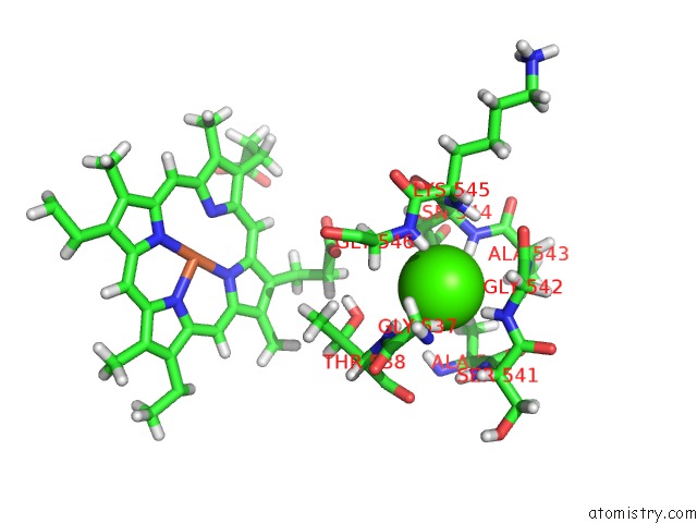
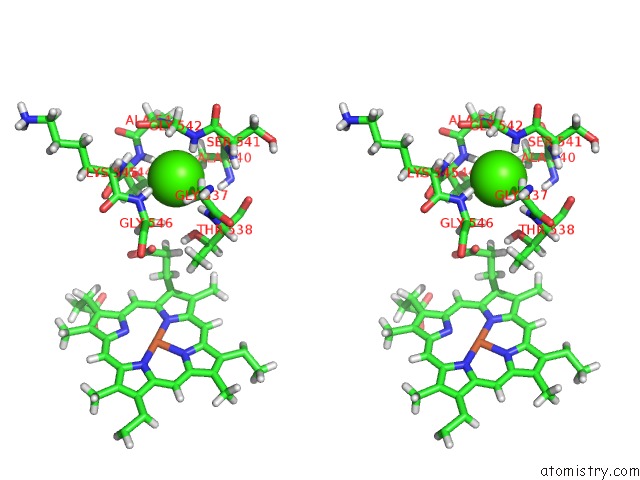
Calcium binding site 3 out of 4 in 6r2q
Go back to
Calcium binding site 3 out
of 4 in the Structure of the Mtr Complex
Mono view
Stereo pair view
Mono view
Stereo pair view
A full contact list of Calcium with other atoms in the Ca binding
site number 3 of Structure of the Mtr Complex within 5.0Å range:
|
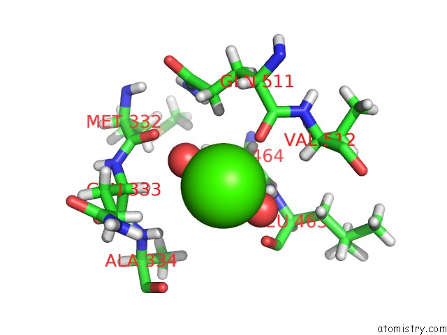
Calcium binding site 4 out of 4 in 6r2q
Go back to
Calcium binding site 4 out
of 4 in the Structure of the Mtr Complex
Mono view
Stereo pair view
Mono view
Stereo pair view
A full contact list of Calcium with other atoms in the Ca binding
site number 4 of Structure of the Mtr Complex within 5.0Å range:
|
Reference:
M.J.Edwards,
G.F.White,
J.N.Butt,
D.J.Richardson,
T.A.Clarke.
The Crystal Structure of A Biological Insulated Transmembrane Molecular Wire. Cell 2020.
ISSN: ISSN 1097-4172
PubMed: 32289252
DOI: 10.1016/J.CELL.2020.03.032
Page generated: Wed Jul 9 17:27:43 2025
ISSN: ISSN 1097-4172
PubMed: 32289252
DOI: 10.1016/J.CELL.2020.03.032
Last articles
Mg in 5SBEMg in 5SBD
Mg in 5SBC
Mg in 5SBB
Mg in 5SBA
Mg in 5SB8
Mg in 5SB9
Mg in 5SB7
Mg in 5SB6
Mg in 5SB4